


Crystal structure of UDP-N-acetylglucosamine 1-carboxyvinyltransferase (UDP-N-acetylglucosamine enolpyruvyl transferase, EPT) from Pseudomonas aeruginosa
Abendroth, J., Dranow, D.M., Lorimer, D.D., Edwards, T.E.To be published.
Experimental Data Snapshot
Starting Model: experimental
View more details
Entity ID: 1 | |||||
|---|---|---|---|---|---|
| Molecule | Chains | Sequence Length | Organism | Details | Image |
| UDP-N-acetylglucosamine 1-carboxyvinyltransferase | 429 | Pseudomonas aeruginosa PAO1 | Mutation(s): 0 Gene Names: murA, PA4450 EC: 2.5.1.7 | 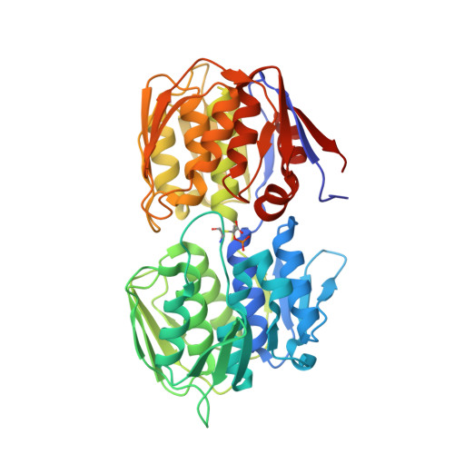 | |
UniProt | |||||
Entity Groups | |||||
| Sequence Clusters | 30% Identity50% Identity70% Identity90% Identity95% Identity100% Identity | ||||
| UniProt Group | Q9HVW7 | ||||
Sequence AnnotationsExpand | |||||
Reference Sequence | |||||
| Ligands 2 Unique | |||||
|---|---|---|---|---|---|
| ID | Chains | Name / Formula / InChI Key | 2D Diagram | 3D Interactions | |
| EPU Download:Ideal Coordinates CCD File | E [auth A], G [auth B], I [auth C], L [auth D] | URIDINE-DIPHOSPHATE-2(N-ACETYLGLUCOSAMINYL) BUTYRIC ACID C20 H29 N3 O19 P2 BEGZZYPUNCJHKP-DBYWSUQTSA-N |  | ||
| MG Download:Ideal Coordinates CCD File | F [auth A] H [auth B] J [auth C] K [auth C] M [auth D] | MAGNESIUM ION Mg JLVVSXFLKOJNIY-UHFFFAOYSA-N |  | ||
| Modified Residues 1 Unique | |||||
|---|---|---|---|---|---|
| ID | Chains | Type | Formula | 2D Diagram | Parent |
| QPA Query on QPA | A, B, C, D | L-PEPTIDE LINKING | C6 H12 N O8 P S |  | CYS |
| Length ( Å ) | Angle ( ˚ ) |
|---|---|
| a = 130.17 | α = 90 |
| b = 166.81 | β = 90 |
| c = 81.45 | γ = 90 |
| Software Name | Purpose |
|---|---|
| XDS | data reduction |
| XSCALE | data scaling |
| PHASER | phasing |
| ARP | model building |
| Coot | model building |
| PHENIX | refinement |


