5BPF | pdb_00005bpf

Crystal structure of ADP complexed D-alanine-D-alanine ligase(DDL) from Yersinia pestis


Experimental Data Snapshot

  • Method: X-RAY DIFFRACTION
  • Resolution: 2.28 Å
  • R-Value Free: 
    0.253 (Depositor), 0.253 (DCC) 
  • R-Value Work: 
    0.186 (Depositor), 0.191 (DCC) 
  • R-Value Observed: 
    0.189 (Depositor) 

Starting Model: experimental
View more details

wwPDB Validation 3D Report Full Report

Validation slider image for 5BPF

Ligand Structure Quality Assessment 


This is version 1.1 of the entry. See complete history

Literature

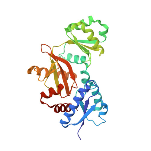
Structure of D-alanine-D-alanine ligase from Yersinia pestis: nucleotide phosphate recognition by the serine loop.

Tran, H.T.Hong, M.K.Ngo, H.P.Huynh, K.H.Ahn, Y.J.Wang, Z.Kang, L.W.

(2016) Acta Crystallogr D Struct Biol 72: 12-21

  • DOI: https://doi.org/10.1107/S2059798315021671
  • Primary Citation Related Structures: 
    4ZQI, 5BPF, 5BPH, 5C1O, 5C1P

  • PubMed Abstract: 

    D-Alanyl-D-alanine is an essential precursor of bacterial peptidoglycan and is synthesized by D-alanine-D-alanine ligase (DDL) with hydrolysis of ATP; this reaction makes DDL an important drug target for the development of antibacterial agents. Five crystal structures of DDL from Yersinia pestis (YpDDL) were determined at 1.7-2.5 Å resolution: apo, AMP-bound, ADP-bound, adenosine 5'-(β,γ-imido)triphosphate-bound, and D-alanyl-D-alanine- and ADP-bound structures. YpDDL consists of three domains, in which four loops, loop 1, loop 2 (the serine loop), loop 3 (the ω-loop) and loop 4, constitute the binding sites for two D-alanine molecules and one ATP molecule. Some of them, especially the serine loop and the ω-loop, show flexible conformations, and the serine loop is mainly responsible for the conformational change in substrate nucleotide phosphates. Enzyme-kinetics assays were carried out for both the D-alanine and ATP substrates and a substrate-binding mechanism was proposed for YpDDL involving conformational changes of the loops.


  • Organizational Affiliation
    • Department of Biological Sciences, Konkuk University, Hwayang-dong, Gwangjin-gu, Seoul 143-701, Republic of Korea.

Macromolecule Content 

  • Total Structure Weight: 134.81 kDa 
  • Atom Count: 9,628 
  • Modeled Residue Count: 1,194 
  • Deposited Residue Count: 1,224 
  • Unique protein chains: 1

Macromolecules

Find similar proteins by:|  3D Structure
Entity ID: 1
MoleculeChains  Sequence LengthOrganismDetailsImage
D-alanine-D-alanine ligase
A, B, C, D
306Yersinia pestisMutation(s): 0 
Gene Names: ddlddlBYPO0557y3624YP_3627
EC: 6.3.2.4
UniProt
Find proteins for Q8ZIE7 (Yersinia pestis)
Explore Q8ZIE7 
Go to UniProtKB:  Q8ZIE7
Entity Groups
Sequence Clusters30% Identity50% Identity70% Identity90% Identity95% Identity100% Identity
UniProt GroupQ8ZIE7
Sequence Annotations
Expand
Reference Sequence

Small Molecules

Ligands 4 Unique
IDChains Name / Formula / InChI Key2D Diagram3D Interactions
ADP

Query on ADP


Download:Ideal Coordinates CCD File
I [auth B],
M [auth C],
R [auth D]
ADENOSINE-5'-DIPHOSPHATE
C10 H15 N5 O10 P2
XTWYTFMLZFPYCI-KQYNXXCUSA-N
GOL

Query on GOL


Download:Ideal Coordinates CCD File
F [auth A],
G [auth A],
Q [auth C],
T [auth D],
U [auth D]
GLYCEROL
C3 H8 O3
PEDCQBHIVMGVHV-UHFFFAOYSA-N
ACT

Query on ACT


Download:Ideal Coordinates CCD File
K [auth B],
L [auth B],
O [auth C],
P [auth C]
ACETATE ION
C2 H3 O2
QTBSBXVTEAMEQO-UHFFFAOYSA-M
NA

Query on NA


Download:Ideal Coordinates CCD File
E [auth A],
H [auth A],
J [auth B],
N [auth C],
S [auth D]
SODIUM ION
Na
FKNQFGJONOIPTF-UHFFFAOYSA-N

Experimental Data & Validation

Experimental Data

  • Method: X-RAY DIFFRACTION
  • Resolution: 2.28 Å
  • R-Value Free:  0.253 (Depositor), 0.253 (DCC) 
  • R-Value Work:  0.186 (Depositor), 0.191 (DCC) 
  • R-Value Observed: 0.189 (Depositor) 
Space Group: P 21 21 21
Unit Cell:
Length ( Å )Angle ( ˚ )
a = 62.872α = 90
b = 106.475β = 90
c = 211.462γ = 90
Software Package:
Software NamePurpose
REFMACrefinement
PDB_EXTRACTdata extraction
DENZOdata reduction
SCALEPACKdata scaling
PHASERphasing

Structure Validation

View Full Validation Report


Ligand Structure Quality Assessment 


Entry History 

Deposition Data

Revision History  (Full details and data files)

  • Version 1.0: 2016-03-02
    Type: Initial release
  • Version 1.1: 2023-11-08
    Changes: Data collection, Database references, Derived calculations, Refinement description