Rotameric preferences of a protein spin label at edge-strand beta-sheet sites.
Cunningham, T.F., Pornsuwan, S., Horne, W.S., Saxena, S.(2016) Protein Sci 25: 1049-1060
- PubMed: 26948069 Search on PubMedSearch on PubMed Central
- DOI: https://doi.org/10.1002/pro.2918
- Primary Citation Related Structures:
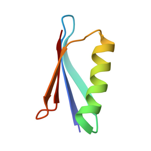
5BMG, 5BMH, 5BMI - PubMed Abstract:
Protein spin labeling to yield the nitroxide-based R1 side chain is a powerful method to measure protein dynamics and structure by electron spin resonance. However, R1 measurements are complicated by the flexibility of the side chain. While analysis approaches for solvent-exposed α-helical environment have been developed to partially account for flexibility, similar work in β-sheets is lacking. The goal of this study is to provide the first essential steps for understanding the conformational preferences of R1 within edge β-strands using X-ray crystallography and double electron electron resonance (DEER) distance measurements. Crystal structures yielded seven rotamers for a non-hydrogen-bonded site and three rotamers for a hydrogen-bonded site. The observed rotamers indicate contextual differences in R1 conformational preferences compared to other solvent-exposed environments. For the DEER measurements, each strand site was paired with the same α-helical site elsewhere on the protein. The most probable distance observed by DEER is rationalized based on the rotamers observed in the crystal structure. Additionally, the appropriateness of common molecular modeling methods that account for R1 conformational preferences are assessed for the β-sheet environment. These results show that interpretation of R1 behavior in β-sheets is difficult and indicate further development is needed for these computational methods to correctly relate DEER distances to protein structure at edge β-strand sites.
- Department of Chemistry, University of Pittsburgh, 219 Parkman Ave, Pittsburgh, Pennsylvania, 15260.
Organizational Affiliation: