5BJU

X-ray structure of the PglF dehydratase from Campylobacter jejuni in complex with UDP and NAD(H)


Experimental Data Snapshot

  • Method: X-RAY DIFFRACTION
  • Resolution: 2.00 Å
  • R-Value Free: 0.237 
  • R-Value Work: 0.193 
  • R-Value Observed: 0.195 

wwPDB Validation   3D Report Full Report


Ligand Structure Quality Assessment 


This is version 1.4 of the entry. See complete history


Literature

Structural and Biochemical Investigation of PglF from Campylobacter jejuni Reveals a New Mechanism for a Member of the Short Chain Dehydrogenase/Reductase Superfamily.

Riegert, A.S.Thoden, J.B.Schoenhofen, I.C.Watson, D.C.Young, N.M.Tipton, P.A.Holden, H.M.

(2017) Biochemistry 56: 6030-6040

  • DOI: https://doi.org/10.1021/acs.biochem.7b00910
  • Primary Citation of Related Structures:  
    5BJU, 5BJV, 5BJW, 5BJX, 5BJY

  • PubMed Abstract: 

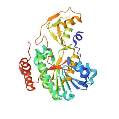
    Within recent years it has become apparent that protein glycosylation is not limited to eukaryotes. Indeed, in Campylobacter jejuni, a Gram-negative bacterium, more than 60 of its proteins are known to be glycosylated. One of the sugars found in such glycosylated proteins is 2,4-diacetamido-2,4,6-trideoxy-α-d-glucopyranose, hereafter referred to as QuiNAc4NAc. The pathway for its biosynthesis, initiating with UDP-GlcNAc, requires three enzymes referred to as PglF, PglE, and PlgD. The focus of this investigation is on PglF, an NAD + -dependent sugar 4,6-dehydratase known to belong to the short chain dehydrogenase/reductase (SDR) superfamily. Specifically, PglF catalyzes the first step in the pathway, namely, the dehydration of UDP-GlcNAc to UDP-2-acetamido-2,6-dideoxy-α-d-xylo-hexos-4-ulose. Most members of the SDR superfamily contain a characteristic signature sequence of YXXXK where the conserved tyrosine functions as a catalytic acid or a base. Strikingly, in PglF, this residue is a methionine. Here we describe a detailed structural and functional investigation of PglF from C. jejuni. For this investigation five X-ray structures were determined to resolutions of 2.0 Å or better. In addition, kinetic analyses of the wild-type and site-directed variants were performed. On the basis of the data reported herein, a new catalytic mechanism for a SDR superfamily member is proposed that does not require the typically conserved tyrosine residue.


  • Organizational Affiliation

    Department of Biochemistry, University of Wisconsin , Madison, Wisconsin 53706, United States.


Macromolecules
Find similar proteins by:  (by identity cutoff)  |  3D Structure
Entity ID: 1
MoleculeChains Sequence LengthOrganismDetailsImage
WlaL protein
A, B
366Campylobacter jejuniMutation(s): 0 
Gene Names: wlaL
UniProt
Find proteins for O86159 (Campylobacter jejuni)
Explore O86159 
Go to UniProtKB:  O86159
Entity Groups  
Sequence Clusters30% Identity50% Identity70% Identity90% Identity95% Identity100% Identity
UniProt GroupO86159
Sequence Annotations
Expand
  • Reference Sequence
Small Molecules
Ligands 4 Unique
IDChains Name / Formula / InChI Key2D Diagram3D Interactions
NAD
Query on NAD

Download Ideal Coordinates CCD File 
D [auth A],
F [auth B]
NICOTINAMIDE-ADENINE-DINUCLEOTIDE
C21 H27 N7 O14 P2
BAWFJGJZGIEFAR-NNYOXOHSSA-N
UDP
Query on UDP

Download Ideal Coordinates CCD File 
C [auth A],
E [auth B]
URIDINE-5'-DIPHOSPHATE
C9 H14 N2 O12 P2
XCCTYIAWTASOJW-XVFCMESISA-N
EDO
Query on EDO

Download Ideal Coordinates CCD File 
G [auth B]1,2-ETHANEDIOL
C2 H6 O2
LYCAIKOWRPUZTN-UHFFFAOYSA-N
NA
Query on NA

Download Ideal Coordinates CCD File 
H [auth B],
I [auth B]
SODIUM ION
Na
FKNQFGJONOIPTF-UHFFFAOYSA-N
Experimental Data & Validation

Experimental Data

  • Method: X-RAY DIFFRACTION
  • Resolution: 2.00 Å
  • R-Value Free: 0.237 
  • R-Value Work: 0.193 
  • R-Value Observed: 0.195 
  • Space Group: P 21 21 21
Unit Cell:
Length ( Å )Angle ( ˚ )
a = 69.135α = 90
b = 107.882β = 90
c = 110.208γ = 90
Software Package:
Software NamePurpose
REFMACrefinement
SAINTdata reduction
SADABSdata scaling
PHASERphasing

Structure Validation

View Full Validation Report


Ligand Structure Quality Assessment 


Entry History & Funding Information

Deposition Data


Funding OrganizationLocationGrant Number
National Institutes of Health/National Institute of General Medical Sciences (NIH/NIGMS)United StatesGM115921

Revision History  (Full details and data files)

  • Version 1.0: 2017-11-08
    Type: Initial release
  • Version 1.1: 2017-11-29
    Changes: Database references
  • Version 1.2: 2017-12-06
    Changes: Author supporting evidence
  • Version 1.3: 2020-01-01
    Changes: Author supporting evidence
  • Version 1.4: 2024-03-06
    Changes: Data collection, Database references, Derived calculations, Refinement description