5B2R | pdb_00005b2r

Crystal structure of the Streptococcus pyogenes Cas9 VQR variant in complex with sgRNA and target DNA (TGA PAM)


Experimental Data Snapshot

  • Method: X-RAY DIFFRACTION
  • Resolution: 2.00 Å
  • R-Value Free: 
    0.230 (Depositor), 0.231 (DCC) 
  • R-Value Work: 
    0.201 (Depositor), 0.203 (DCC) 
  • R-Value Observed: 
    0.202 (Depositor) 

Starting Model: experimental
View more details

wwPDB Validation 3D Report Full Report

Validation slider image for 5B2R

This is version 1.3 of the entry. See complete history

Literature

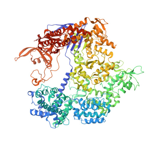
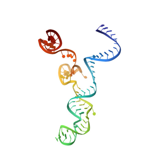
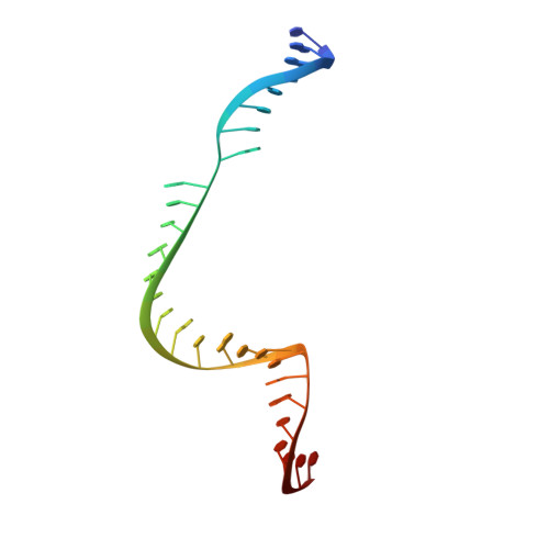
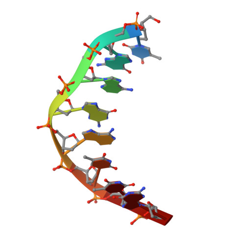
Structural Basis for the Altered PAM Specificities of Engineered CRISPR-Cas9

Hirano, S.Nishimasu, H.Ishitani, R.Nureki, O.

(2016) Mol Cell 61: 886-894

  • DOI: https://doi.org/10.1016/j.molcel.2016.02.018
  • Primary Citation Related Structures: 
    5B2R, 5B2S, 5B2T

  • PubMed Abstract: 

    The RNA-guided endonuclease Cas9 cleaves double-stranded DNA targets bearing a PAM (protospacer adjacent motif) and complementarity to the guide RNA. A recent study showed that, whereas wild-type Streptococcus pyogenes Cas9 (SpCas9) recognizes the 5'-NGG-3' PAM, the engineered VQR, EQR, and VRER SpCas9 variants recognize the 5'-NGA-3', 5'-NGAG-3', and 5'-NGCG-3' PAMs, respectively, thus expanding the targetable sequences in Cas9-mediated genome editing applications. Here, we present the high-resolution crystal structures of the three SpCas9 variants in complexes with a single-guide RNA and its altered PAM-containing, partially double-stranded DNA targets. A structural comparison of the three SpCas9 variants with wild-type SpCas9 revealed that the multiple mutations synergistically induce an unexpected displacement in the phosphodiester backbone of the PAM duplex, thereby allowing the SpCas9 variants to directly recognize the altered PAM nucleotides. Our findings explain the altered PAM specificities of the SpCas9 variants and establish a framework for further rational engineering of CRISPR-Cas9.


  • Organizational Affiliation
    • Department of Biological Sciences, Graduate School of Science, The University of Tokyo, 2-11-16 Yayoi, Bunkyo-ku, Tokyo 113-0032, Japan.

Macromolecule Content 

  • Total Structure Weight: 196.93 kDa 
  • Atom Count: 13,799 
  • Modeled Residue Count: 1,436 
  • Deposited Residue Count: 1,489 
  • Unique protein chains: 1
  • Unique nucleic acid chains: 3

Macromolecules


Find similar proteins by:|  3D Structure
Entity ID: 2
MoleculeChains  Sequence LengthOrganismDetailsImage
CRISPR-associated endonuclease Cas91,372Streptococcus pyogenes serotype M1Mutation(s): 7 
Gene Names: cas9csn1SPy_1046
EC: 3.1
UniProt
Find proteins for Q99ZW2 (Streptococcus pyogenes serotype M1)
Explore Q99ZW2 
Go to UniProtKB:  Q99ZW2
Entity Groups
Sequence Clusters30% Identity50% Identity70% Identity90% Identity95% Identity100% Identity
UniProt GroupQ99ZW2
Sequence Annotations
Expand
Reference Sequence
Find similar nucleic acids by:  (by identity cutoff) 
Entity ID: 1
MoleculeChains LengthOrganismImage
Guide RNA81Streptococcus pyogenes
Sequence Annotations
Expand
Reference Sequence
Find similar nucleic acids by:  (by identity cutoff) 
Entity ID: 3
MoleculeChains LengthOrganismImage
Target DNA28Streptococcus pyogenes
Sequence Annotations
Expand
Reference Sequence
Find similar nucleic acids by:  Sequence
Entity ID: 4
MoleculeChains LengthOrganismImage
Non-target DNA, DNA (5'-D(*TP*GP*AP*GP*AP*TP*TP*G)-3')8Streptococcus pyogenes
Sequence Annotations
Expand
Reference Sequence

Small Molecules

Ligands 4 Unique
IDChains Name / Formula / InChI Key2D Diagram3D Interactions
EDO

Query on EDO


Download:Ideal Coordinates CCD File
J [auth A],
U [auth B],
V [auth B],
W [auth B]
1,2-ETHANEDIOL
C2 H6 O2
LYCAIKOWRPUZTN-UHFFFAOYSA-N
ACT

Query on ACT


Download:Ideal Coordinates CCD File
K [auth A]ACETATE ION
C2 H3 O2
QTBSBXVTEAMEQO-UHFFFAOYSA-M
K

Query on K


Download:Ideal Coordinates CCD File
E [auth A]
F [auth A]
G [auth A]
L [auth A]
M [auth B]
E [auth A],
F [auth A],
G [auth A],
L [auth A],
M [auth B],
N [auth B],
O [auth B],
P [auth B],
Q [auth B],
R [auth B],
S [auth B]
POTASSIUM ION
K
NPYPAHLBTDXSSS-UHFFFAOYSA-N
MG

Query on MG


Download:Ideal Coordinates CCD File
H [auth A],
I [auth A],
T [auth B]
MAGNESIUM ION
Mg
JLVVSXFLKOJNIY-UHFFFAOYSA-N

Experimental Data & Validation

Experimental Data

  • Method: X-RAY DIFFRACTION
  • Resolution: 2.00 Å
  • R-Value Free:  0.230 (Depositor), 0.231 (DCC) 
  • R-Value Work:  0.201 (Depositor), 0.203 (DCC) 
  • R-Value Observed: 0.202 (Depositor) 
Space Group: C 1 2 1
Unit Cell:
Length ( Å )Angle ( ˚ )
a = 177.764α = 90
b = 67.744β = 111.23
c = 187.594γ = 90
Software Package:
Software NamePurpose
Aimlessdata scaling
PHENIXrefinement
PDB_EXTRACTdata extraction

Structure Validation

View Full Validation Report


Entry History 

Deposition Data

Revision History  (Full details and data files)

  • Version 1.0: 2016-03-23
    Type: Initial release
  • Version 1.1: 2016-04-06
    Changes: Database references
  • Version 1.2: 2017-10-04
    Changes: Data collection, Database references, Derived calculations
  • Version 1.3: 2023-11-08
    Changes: Data collection, Database references, Derived calculations, Refinement description