5AXG | pdb_00005axg

Crystal structure of thermophilic dextranase from Thermoanaerobacter pseudethanolicus


Experimental Data Snapshot

  • Method: X-RAY DIFFRACTION
  • Resolution: 1.85 Å
  • R-Value Free: 
    0.190 (Depositor), 0.199 (DCC) 
  • R-Value Work: 
    0.166 (Depositor), 0.175 (DCC) 
  • R-Value Observed: 
    0.167 (Depositor) 

Starting Model: experimental
View more details

wwPDB Validation 3D Report Full Report

Validation slider image for 5AXG

Ligand Structure Quality Assessment 


This is version 1.3 of the entry. See complete history

Literature

Crystal structure of thermophilic dextranase from Thermoanaerobacter pseudethanolicus

Suzuki, N.Kishine, N.Fujimoto, Z.Sakurai, M.Momma, M.Ko, J.A.Nam, S.H.Kimura, A.Kim, Y.M.

(2016) J Biochem 159: 331-339

  • DOI: https://doi.org/10.1093/jb/mvv104
  • Primary Citation Related Structures: 
    5AXG, 5AXH

  • PubMed Abstract: 

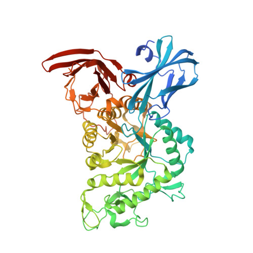
    The crystal structures of the wild type and catalytic mutant Asp-312→Gly in complex with isomaltohexaose of endo-1,6-dextranase from the thermophilic bacterium Thermoanaerobacter pseudethanolicus (TpDex), belonging to the glycoside hydrolase family 66, were determined. TpDex consists of three structural domains, a catalytic domain comprising an (β/α)8-barrel and two β-domains located at both N- and C-terminal ends. The isomaltohexaose-complex structure demonstrated that the isomaltohexaose molecule was bound across the catalytic site, showing that TpDex had six subsites (-4 to +2) in the catalytic cleft. Marked movement of the Trp-376 side-chain along with loop 6, which was the side wall component of the cleft at subsite +1, was observed to occupy subsite +1, indicating that it might expel the cleaved aglycone subsite after the hydrolysis reaction. Structural comparison with other mesophilic enzymes indicated that several structural features of TpDex, loop deletion, salt bridge and surface-exposed charged residue, may contribute to thermostability.


  • Organizational Affiliation
    • Biomolecular Research Unit, National Institute of Agrobiological Sciences, 2-1-2 Kannondai, Tsukuba 305-8602, Japan; zui@affrc.go.jp u9897854@jnu.ac.kr.

Macromolecule Content 

  • Total Structure Weight: 144.35 kDa 
  • Atom Count: 10,614 
  • Modeled Residue Count: 1,167 
  • Deposited Residue Count: 1,236 
  • Unique protein chains: 1

Macromolecules

Find similar proteins by:|  3D Structure
Entity ID: 1
MoleculeChains  Sequence LengthOrganismDetailsImage
Dextranase
A, B
618Thermoanaerobacter pseudethanolicus ATCC 33223Mutation(s): 0 
Gene Names: Teth39_0264
EC: 3.2.1.11
UniProt
Find proteins for B0KBZ7 (Thermoanaerobacter pseudethanolicus (strain ATCC 33223 / 39E))
Explore B0KBZ7 
Go to UniProtKB:  B0KBZ7
Entity Groups
Sequence Clusters30% Identity50% Identity70% Identity90% Identity95% Identity100% Identity
UniProt GroupB0KBZ7
Sequence Annotations
Expand
Reference Sequence

Small Molecules

Ligands 4 Unique
IDChains Name / Formula / InChI Key2D Diagram3D Interactions
CXS

Query on CXS


Download:Ideal Coordinates CCD File
M [auth B]3-CYCLOHEXYL-1-PROPYLSULFONIC ACID
C9 H19 N O3 S
PJWWRFATQTVXHA-UHFFFAOYSA-N
PO4

Query on PO4


Download:Ideal Coordinates CCD File
C [auth A]
D [auth A]
E [auth A]
F [auth A]
G [auth A]
C [auth A],
D [auth A],
E [auth A],
F [auth A],
G [auth A],
N [auth B],
O [auth B],
P [auth B]
PHOSPHATE ION
O4 P
NBIIXXVUZAFLBC-UHFFFAOYSA-K
EDO

Query on EDO


Download:Ideal Coordinates CCD File
H [auth A]
I [auth A]
J [auth A]
K [auth A]
Q [auth B]
H [auth A],
I [auth A],
J [auth A],
K [auth A],
Q [auth B],
R [auth B],
S [auth B],
T [auth B],
U [auth B]
1,2-ETHANEDIOL
C2 H6 O2
LYCAIKOWRPUZTN-UHFFFAOYSA-N
K

Query on K


Download:Ideal Coordinates CCD File
L [auth B]POTASSIUM ION
K
NPYPAHLBTDXSSS-UHFFFAOYSA-N

Experimental Data & Validation

Experimental Data

  • Method: X-RAY DIFFRACTION
  • Resolution: 1.85 Å
  • R-Value Free:  0.190 (Depositor), 0.199 (DCC) 
  • R-Value Work:  0.166 (Depositor), 0.175 (DCC) 
  • R-Value Observed: 0.167 (Depositor) 
Space Group: P 21 21 21
Unit Cell:
Length ( Å )Angle ( ˚ )
a = 81.342α = 90
b = 98.992β = 90
c = 171.13γ = 90
Software Package:
Software NamePurpose
REFMACrefinement
HKL-2000data reduction
SCALEPACKdata scaling
MOLREPphasing

Structure Validation

View Full Validation Report


Ligand Structure Quality Assessment 


Entry History 

& Funding Information

Deposition Data


Funding OrganizationLocationGrant Number
Ministry of Agriculture, Forestry and FisheriesJapan25001A
National Research Foundation of KoreaKorea, Republic Of2010-0020141

Revision History  (Full details and data files)

  • Version 1.0: 2015-11-11
    Type: Initial release
  • Version 1.1: 2016-03-09
    Changes: Database references
  • Version 1.2: 2020-02-26
    Changes: Data collection, Derived calculations
  • Version 1.3: 2023-11-08
    Changes: Data collection, Database references, Derived calculations, Refinement description