Homo-trimeric Structure of the Type IVb Minor Pilin CofB Suggests Mechanism of CFA/III Pilus Assembly in Human Enterotoxigenic Escherichia coli
Kawahara, K., Oki, H., Fukakusa, S., Yoshida, T., Imai, T., Maruno, T., Kobayashi, Y., Motooka, D., Iida, T., Ohkubo, T., Nakamura, S.(2016) J Mol Biol 428: 1209-1226
- PubMed: 26876601 Search on PubMed
- DOI: https://doi.org/10.1016/j.jmb.2016.02.003
- Primary Citation Related Structures:
5AX6 - PubMed Abstract:
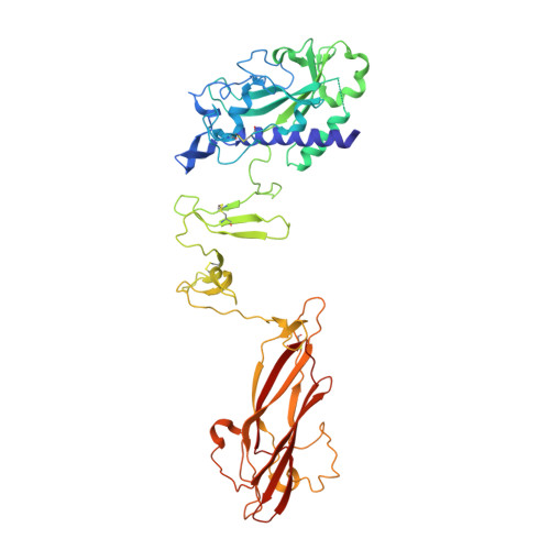
In gram-negative bacteria, the assembly of type IV pilus (T4P) and the evolutionally related pseudopilus of type II secretion system involves specialized structural proteins called pilins and pseudopilins, respectively, and is dynamically regulated to promote bacterial pathogenesis. Previous studies have suggested that a structural "tip"-like hetero-complex formed through the interaction of at least three minor (pseudo) pilins plays an important role in this process, while some members of the pathogenic type IVb subfamily are known to have only one such minor pilin subunit whose function is still unknown. Here, we determined the crystal structure of the type IVb minor pilin CofB of colonization factor antigen/III from human enterotoxigenic Escherichia coli at 1.88-Å resolution. The crystal structure, in conjunction with physicochemical analysis in solution, reveals a symmetrical homo-trimeric arrangement distinct from the hetero-complexes of minor (pseudo) pilins observed in other T4P and type II secretion systems. Each CofB monomer adopts a unique three-domain architecture, in which the C-terminal β-sheet-rich lectin domain can effectively initiate trimer association of its pilin-like N-terminal domain through extensive hydrophobic interactions followed by domain swapping at the central hinge-like domain. Deletion of cofB produces a phenotype with no detectable pili formation on the cell surface, while molecular modeling indicates that the characteristic homo-trimeric structure of CofB is well situated at the pilus tip of colonization factor antigen/III formed by the major pilin CofA, suggesting a role for the minor pilin in the efficient initiation of T4P assembly.
- Graduate School of Pharmaceutical Sciences, Osaka University, 1-6 Yamadaoka, Suita, Osaka 565-0871, Japan.
Organizational Affiliation: