5AVE | pdb_00005ave

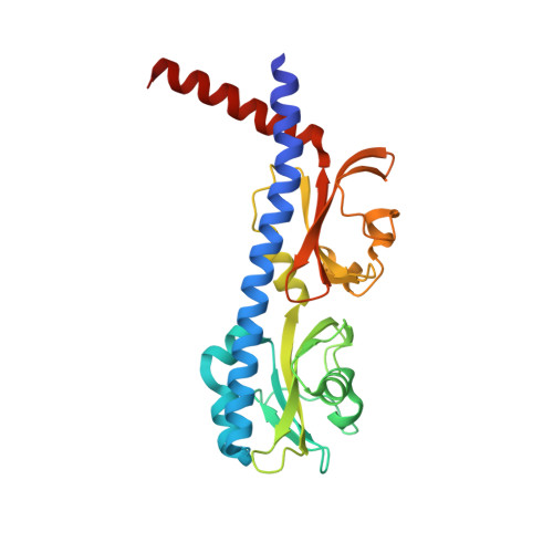
The ligand binding domain of Mlp37 with serine


Experimental Data Snapshot

  • Method: X-RAY DIFFRACTION
  • Resolution: 1.80 Å
  • R-Value Free: 
    0.214 (Depositor), 0.227 (DCC) 
  • R-Value Work: 
    0.188 (Depositor) 
  • R-Value Observed: 
    0.189 (Depositor) 

wwPDB Validation 3D Report Full Report

Validation slider image for 5AVE

This is version 1.2 of the entry. See complete history. 

Literature

Identification of a Vibrio cholerae chemoreceptor that senses taurine and amino acids as attractants

Nishiyama, S., Takahashi, Y., Yamamoto, K., Suzuki, D., Itoh, Y., Sumita, K., Uchida, Y., Homma, M., Imada, K., Kawagishi, I.

(2016) Sci Rep 6: 20866-20866

  • DOI: https://doi.org/10.1038/srep20866
  • Primary Citation Related Structures: 
    5AVE, 5AVF

  • PubMed Abstract: 

    Vibrio cholerae, the etiological agent of cholera, was found to be attracted by taurine (2-aminoethanesulfonic acid), a major constituent of human bile. Mlp37, the closest homolog of the previously identified amino acid chemoreceptor Mlp24, was found to mediate taxis to taurine as well as L-serine, L-alanine, L-arginine, and other amino acids. Methylation of Mlp37 was enhanced upon the addition of taurine and amino acids. Isothermal titration calorimetry demonstrated that a purified periplasmic fragment of Mlp37 binds directly to taurine, L-serine, L-alanine and L-arginine. Crystal structures of the periplamic domain of Mlp37 revealed that L-serine and taurine bind to the membrane-distal PAS domain in essentially in the same way. The structural information was supported by characterising the in vivo properties of alanine-substituted mutant forms of Mlp37. The fact that the ligand-binding domain of the L-serine complex had a small opening, which would accommodate a larger R group, accounts for the broad ligand specificity of Mlp37 and allowed us to visualise ligand binding to Mlp37 with fluorescently labelled L-serine. Taken together, we conclude that Mlp37 serves as the major chemoreceptor for taurine and various amino acids.


  • Organizational Affiliation: 
    • Department of Frontier Bioscience, Hosei University, Kajino-cho, Koganei, Tokyo 184-8584, Japan.

Macromolecule Content 

  • Total Structure Weight: 28.27 kDa 
  • Atom Count: 2,185 
  • Modeled Residue Count: 252 
  • Deposited Residue Count: 257 
  • Unique protein chains: 1

Macromolecules

Find similar proteins by:|  3D Structure
Entity ID: 1
MoleculeChains  Sequence LengthOrganismDetailsImage
Methyl-accepting chemotaxis (MCP) signaling domain protein257Vibrio choleraeMutation(s): 0 
Gene Names: DN30_191
UniProt
Find proteins for A0A0H3AES3 (Vibrio cholerae serotype O1 (strain ATCC 39541 / Classical Ogawa 395 / O395))
Explore A0A0H3AES3 
Go to UniProtKB:  A0A0H3AES3
Entity Groups
Sequence Clusters30% Identity50% Identity70% Identity90% Identity95% Identity100% Identity
UniProt GroupA0A0H3AES3
Sequence Annotations
Expand
Reference Sequence

Small Molecules

Ligands 1 Unique
IDChains Name / Formula / InChI Key2D Diagram3D Interactions
SER

Query on SER


Download:Ideal Coordinates CCD File
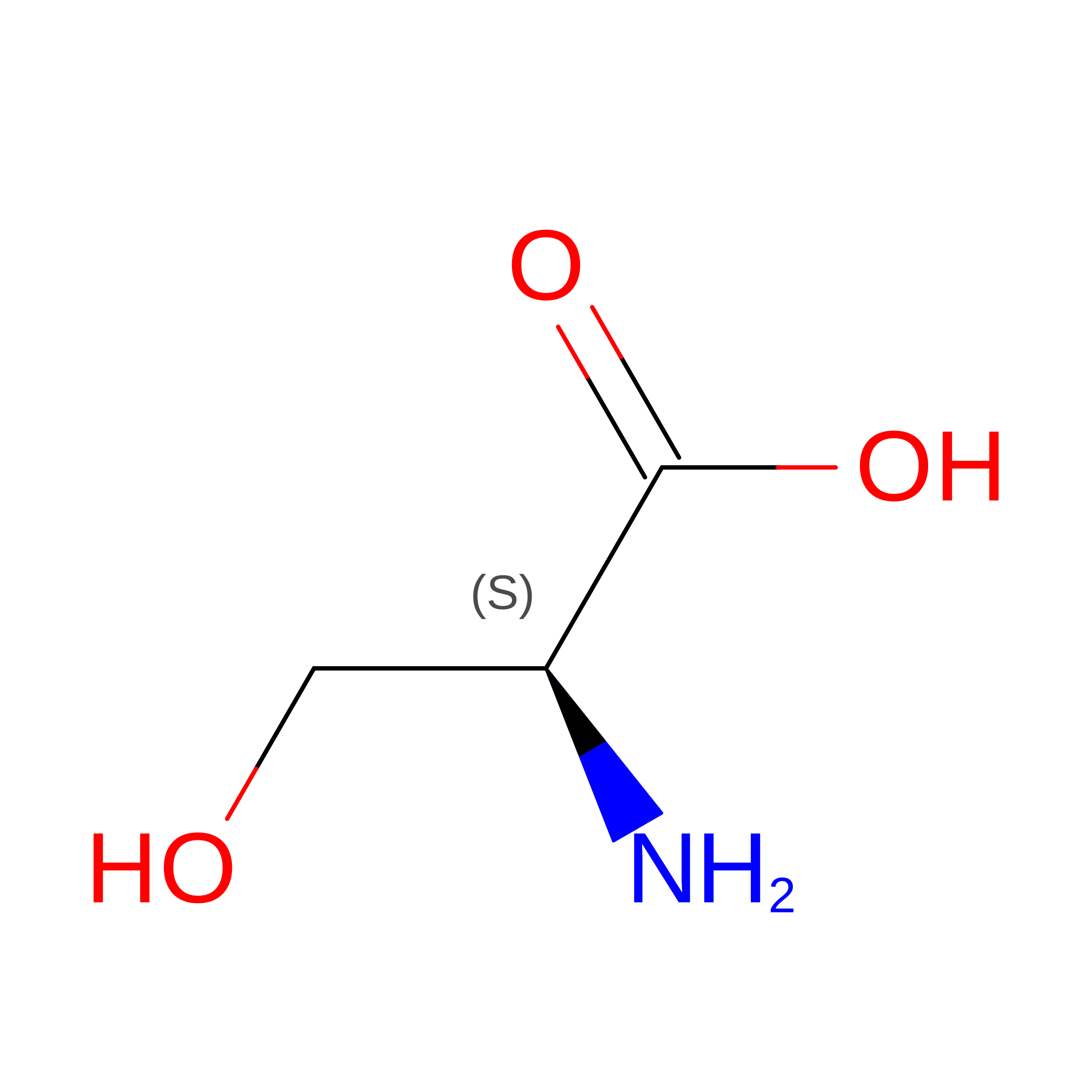
B [auth A]SERINE
C3 H7 N O3
MTCFGRXMJLQNBG-REOHCLBHSA-N

Experimental Data & Validation

Experimental Data

  • Method: X-RAY DIFFRACTION
  • Resolution: 1.80 Å
  • R-Value Free:  0.214 (Depositor), 0.227 (DCC) 
  • R-Value Work:  0.188 (Depositor) 
  • R-Value Observed: 0.189 (Depositor) 
Space Group: C 2 2 21
Unit Cell:
Length ( Å )Angle ( ˚ )
a = 98.72α = 90
b = 134.22β = 90
c = 50.53γ = 90
Software Package:
Software NamePurpose
PHENIXrefinement
MOSFLMdata reduction
SCALAdata scaling
PHENIXphasing

Structure Validation

View Full Validation Report


Entry History 

& Funding Information

Deposition Data


Funding OrganizationLocationGrant Number
Japan--

Revision History  (Full details and data files)

  • Version 1.0: 2016-03-02
    Type: Initial release
  • Version 1.1: 2020-02-19
    Changes: Data collection, Derived calculations
  • Version 1.2: 2024-03-20
    Changes: Data collection, Database references