5AUT

Crystal structure of DAPK1 in complex with ANS.


Experimental Data Snapshot

  • Method: X-RAY DIFFRACTION
  • Resolution: 1.70 Å
  • R-Value Free: 0.202 
  • R-Value Work: 0.170 
  • R-Value Observed: 0.172 

Starting Model: experimental
View more details

wwPDB Validation   3D Report Full Report


Ligand Structure Quality Assessment 


This is version 1.2 of the entry. See complete history


Literature

Structural Insight into the Interactions between Death-Associated Protein Kinase 1 and Natural Flavonoids.

Yokoyama, T.Kosaka, Y.Mizuguchi, M.

(2015) J Med Chem 58: 7400-7408

  • DOI: https://doi.org/10.1021/acs.jmedchem.5b00893
  • Primary Citation of Related Structures:  
    5AUT, 5AUU, 5AUV, 5AUW, 5AUX, 5AUY, 5AUZ, 5AV0, 5AV1, 5AV2, 5AV3, 5AV4

  • PubMed Abstract: 

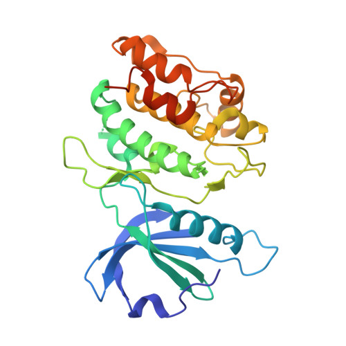
    Death-associated protein kinase 1 (DAPK1) is a 160 kDa serine/threonine protein kinase that belongs to the Ca(2+)/calmodulin-dependent protein kinase subfamily. DAPK1 is a possible target for the treatment of acute ischemic stroke and endometrial adenocarcinomas. In the present study, we investigated the binding characteristics of 17 natural flavonoids to DAPK1 using a 1-anilinonaphthalene-8-sulfonic acid competitive binding assay and revealed that morin was the strongest binder among the selected compounds. The crystallographic analysis of DAPK1 and 7 selected flavonoid complexes revealed the structure-binding affinity relationship in atomic-level detail. It was suggested that the high affinity of morin could be accounted for by the ionic interaction between 2'-OH and K42 and that such an interaction would not take place with either cyclin-dependent protein kinases or PIM kinases because of their broader entrance regions. Thus, morin would be a more selective inhibitor of DAPK1 than either of these other types of kinases. In addition, we found that the binding of kaempferol to DAPK1 was associated with a chloride ion. The present study provides a better understanding of the molecular properties of the ATP site of DAPK1 and may be useful for the design of specific DAPK1 inhibitors.


  • Organizational Affiliation

    Faculty of Pharmaceutical Sciences, University of Toyama , 2630 Sugitani, Toyama 930-0914, Japan.


Macromolecules
Find similar proteins by:  (by identity cutoff)  |  3D Structure
Entity ID: 1
MoleculeChains Sequence LengthOrganismDetailsImage
Death-associated protein kinase 1293Homo sapiensMutation(s): 0 
Gene Names: DAPK1DAPK
EC: 2.7.11.1
UniProt & NIH Common Fund Data Resources
Find proteins for P53355 (Homo sapiens)
Explore P53355 
Go to UniProtKB:  P53355
GTEx:  ENSG00000196730 
Entity Groups  
Sequence Clusters30% Identity50% Identity70% Identity90% Identity95% Identity100% Identity
UniProt GroupP53355
Sequence Annotations
Expand
  • Reference Sequence
Small Molecules
Ligands 1 Unique
IDChains Name / Formula / InChI Key2D Diagram3D Interactions
2AN
Query on 2AN

Download Ideal Coordinates CCD File 
B [auth A]8-ANILINO-1-NAPHTHALENE SULFONATE
C16 H13 N O3 S
FWEOQOXTVHGIFQ-UHFFFAOYSA-N
Experimental Data & Validation

Experimental Data

  • Method: X-RAY DIFFRACTION
  • Resolution: 1.70 Å
  • R-Value Free: 0.202 
  • R-Value Work: 0.170 
  • R-Value Observed: 0.172 
  • Space Group: P 21 21 21
Unit Cell:
Length ( Å )Angle ( ˚ )
a = 46.774α = 90
b = 62.26β = 90
c = 88.6γ = 90
Software Package:
Software NamePurpose
PHENIXrefinement
HKL-2000data reduction
SCALEPACKdata scaling
PHENIXphasing

Structure Validation

View Full Validation Report


Ligand Structure Quality Assessment 


Entry History 

Deposition Data

Revision History  (Full details and data files)

  • Version 1.0: 2015-10-07
    Type: Initial release
  • Version 1.1: 2020-02-19
    Changes: Data collection, Database references, Derived calculations, Source and taxonomy, Structure summary
  • Version 1.2: 2023-11-08
    Changes: Data collection, Database references, Refinement description