Terahertz Radiation Induces Non-Thermal Structural Changes Associated with Frohlich Condensation in a Protein Crystal
Lundholm, I., Rodilla, H., Wahlgren, W.Y., Duelli, A., Bourenkov, G., Vukusic, J., Friedman, R., Stake, J., Schneider, T., Katona, G.(2015) Struct Dyn 2: 54702
- PubMed: 26798828 Search on PubMedSearch on PubMed Central
- DOI: https://doi.org/10.1063/1.4931825
- Primary Citation Related Structures:
5APC, 5APD, 5APE, 5APF - PubMed Abstract:
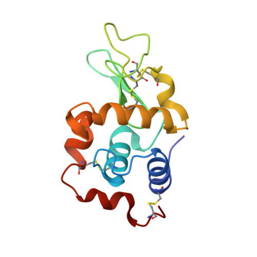
Whether long-range quantum coherent states could exist in biological systems, and beyond low-temperature regimes where quantum physics is known to be applicable, has been the subject to debate for decades. It was proposed by Fröhlich that vibrational modes within protein molecules can order and condense into a lowest-frequency vibrational mode in a process similar to Bose-Einstein condensation, and thus that macroscopic coherence could potentially be observed in biological systems. Despite the prediction of these so-called Fröhlich condensates almost five decades ago, experimental evidence thereof has been lacking. Here, we present the first experimental observation of Fröhlich condensation in a protein structure. To that end, and to overcome the challenges associated with probing low-frequency molecular vibrations in proteins (which has hampered understanding of their role in proteins' function), we combined terahertz techniques with a highly sensitive X-ray crystallographic method to visualize low-frequency vibrational modes in the protein structure of hen-egg white lysozyme. We found that 0.4 THz electromagnetic radiation induces non-thermal changes in electron density. In particular, we observed a local increase of electron density in a long α-helix motif consistent with a subtle longitudinal compression of the helix. These observed electron density changes occur at a low absorption rate indicating that thermalization of terahertz photons happens on a micro- to milli-second time scale, which is much slower than the expected nanosecond time scale due to damping of delocalized low frequency vibrations. Our analyses show that the micro- to milli-second lifetime of the vibration can only be explained by Fröhlich condensation, a phenomenon predicted almost half a century ago, yet never experimentally confirmed.
- Department of Chemistry and Molecular Biology, University of Gothenburg , Gothenburg, Sweden.
Organizational Affiliation: