X-Ray Crystallography-Promoted Drug Design of Carbonic Anhydrase Inhibitors.
Ivanova, J., Leitans, J., Tanc, M., Kazaks, A., Zalubovskis, R., Supuran, C.T., Tars, K.(2015) Chem Commun (Camb) 51: 7108
- PubMed: 25813715 Search on PubMed
- DOI: https://doi.org/10.1039/c5cc01854d
- Primary Citation Related Structures:
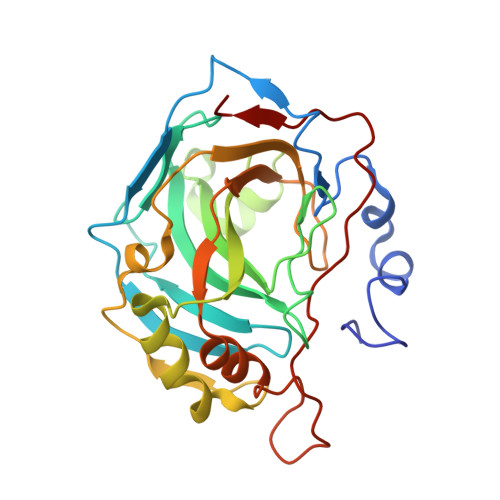
5AMD, 5AMG, 5AML - PubMed Abstract:
1-N-Alkylated-6-sulfamoyl saccharin derivatives were prepared and assayed as carbonic anhydrase inhibitors (CAIs). During X-ray crystallographic experiments an unexpected hydrolysis of the isothiazole ring was evidenced which allowed us to prepare highly potent enzyme inhibitors with selectivity for some isoforms with medical applications.
- Latvian Institute of Organic Synthesis, Aizkraukles 21, LV-1006 Riga, Latvia. raivis@osi.lv.
Organizational Affiliation: