5AM9

Crystal structure of the Angiotensin-1 converting enzyme N-domain in complex with amyloid-beta 10-16


Experimental Data Snapshot

  • Method: X-RAY DIFFRACTION
  • Resolution: 1.80 Å
  • R-Value Free: 0.229 
  • R-Value Work: 0.197 
  • R-Value Observed: 0.198 

wwPDB Validation   3D Report Full Report


This is version 2.1 of the entry. See complete history


Literature

The Kinetic and Structural Characterisation of Amyloid-Beta Metabolism by Human Angiotensin-1- Converting Enzyme (Ace)

Larmuth, K.M.Masuyer, G.Douglas, R.G.Sturrock, E.D.Acharya, K.R.

(2016) FEBS J 283: 1060

  • DOI: https://doi.org/10.1111/febs.13647
  • Primary Citation of Related Structures:  
    5AM8, 5AM9, 5AMA, 5AMB, 5AMC

  • PubMed Abstract: 

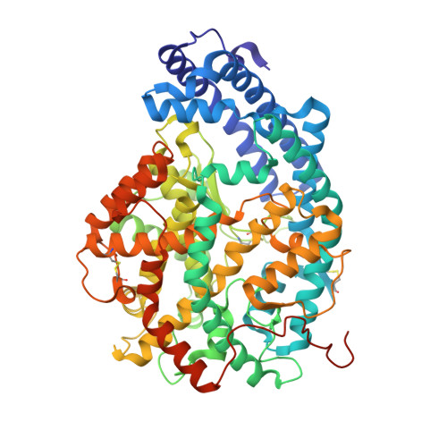
    Angiotensin-1-converting enzyme (ACE), a zinc metallopeptidase, consists of two homologous catalytic domains (N and C) with different substrate specificities. Here we report kinetic parameters of five different forms of human ACE with various amyloid beta (Aβ) substrates together with high resolution crystal structures of the N-domain in complex with Aβ fragments. For the physiological Aβ(1-16) peptide, a novel ACE cleavage site was found at His14-Gln15. Furthermore, Aβ(1-16) was preferentially cleaved by the individual N-domain; however, the presence of an inactive C-domain in full-length somatic ACE (sACE) greatly reduced enzyme activity and affected apparent selectivity. Two fluorogenic substrates, Aβ(4-10)Q and Aβ(4-10)Y, underwent endoproteolytic cleavage at the Asp7-Ser8 bond with all ACE constructs showing greater catalytic efficiency for Aβ(4-10)Y. Surprisingly, in contrast to Aβ(1-16) and Aβ(4-10)Q, sACE showed positive domain cooperativity and the double C-domain (CC-sACE) construct no cooperativity towards Aβ(4-10)Y. The structures of the Aβ peptide-ACE complexes revealed a common mode of peptide binding for both domains which principally targets the C-terminal P2' position to the S2' pocket and recognizes the main chain of the P1' peptide. It is likely that N-domain selectivity for the amyloid peptide is conferred through the N-domain specific S2' residue Thr358. Additionally, the N-domain can accommodate larger substrates through movement of the N-terminal helices, as suggested by the disorder of the hinge region in the crystal structures. Our findings are important for the design of domain selective inhibitors as the differences in domain selectivity are more pronounced with the truncated domains compared to the more physiological full-length forms.


  • Organizational Affiliation

    Department of Integrative Biomedical Sciences, Institute of Infectious Disease and Molecular Medicine, University of Cape Town, South Africa.


Macromolecules
Find similar proteins by:  (by identity cutoff)  |  3D Structure
Entity ID: 1
MoleculeChains Sequence LengthOrganismDetailsImage
ANGIOTENSIN-CONVERTING ENZYME
A, B, C, D
629Homo sapiensMutation(s): 9 
EC: 3.2.1 (PDB Primary Data), 3.4.15.1 (PDB Primary Data)
UniProt & NIH Common Fund Data Resources
Find proteins for P12821 (Homo sapiens)
Explore P12821 
Go to UniProtKB:  P12821
PHAROS:  P12821
GTEx:  ENSG00000159640 
Entity Groups  
Sequence Clusters30% Identity50% Identity70% Identity90% Identity95% Identity100% Identity
UniProt GroupP12821
Sequence Annotations
Expand
  • Reference Sequence
Oligosaccharides

Help

Entity ID: 2
MoleculeChains Length2D Diagram Glycosylation3D Interactions
beta-L-fucopyranose-(1-6)-2-acetamido-2-deoxy-beta-D-glucopyranose
E
2N-Glycosylation
Glycosylation Resources
GlyTouCan:  G61843VN
GlyCosmos:  G61843VN
GlyGen:  G61843VN
Entity ID: 3
MoleculeChains Length2D Diagram Glycosylation3D Interactions
2-acetamido-2-deoxy-beta-D-glucopyranose-(1-4)-2-acetamido-2-deoxy-beta-D-glucopyranose
F, I, L, O
2N-Glycosylation
Glycosylation Resources
GlyTouCan:  G42666HT
GlyCosmos:  G42666HT
GlyGen:  G42666HT
Entity ID: 4
MoleculeChains Length2D Diagram Glycosylation3D Interactions
alpha-D-mannopyranose-(1-4)-2-acetamido-2-deoxy-beta-D-glucopyranose-(1-4)-2-acetamido-2-deoxy-beta-D-glucopyranose
G, J
3N-Glycosylation
Glycosylation Resources
GlyTouCan:  G62182OO
GlyCosmos:  G62182OO
GlyGen:  G62182OO
Entity ID: 5
MoleculeChains Length2D Diagram Glycosylation3D Interactions
alpha-L-fucopyranose-(1-6)-2-acetamido-2-deoxy-beta-D-glucopyranose
H, K, N
2N-Glycosylation
Glycosylation Resources
GlyTouCan:  G86851RC
GlyCosmos:  G86851RC
GlyGen:  G86851RC
Entity ID: 6
MoleculeChains Length2D Diagram Glycosylation3D Interactions
beta-D-mannopyranose-(1-4)-2-acetamido-2-deoxy-beta-D-glucopyranose-(1-4)-[alpha-L-fucopyranose-(1-6)]2-acetamido-2-deoxy-beta-D-glucopyranose
M, P
4N-Glycosylation
Glycosylation Resources
GlyTouCan:  G32152BH
GlyCosmos:  G32152BH
GlyGen:  G32152BH
Small Molecules
Ligands 10 Unique
IDChains Name / Formula / InChI Key2D Diagram3D Interactions
P6G
Query on P6G

Download Ideal Coordinates CCD File 
GA [auth B],
HA [auth B],
X [auth A],
YA [auth D]
HEXAETHYLENE GLYCOL
C12 H26 O7
IIRDTKBZINWQAW-UHFFFAOYSA-N
LYS
Query on LYS

Download Ideal Coordinates CCD File 
R [auth A],
TA [auth D]
LYSINE
C6 H15 N2 O2
KDXKERNSBIXSRK-YFKPBYRVSA-O
GLU
Query on GLU

Download Ideal Coordinates CCD File 
JA [auth C],
Z [auth B]
GLUTAMIC ACID
C5 H9 N O4
WHUUTDBJXJRKMK-VKHMYHEASA-N
GLN
Query on GLN

Download Ideal Coordinates CCD File 
Q [auth A],
SA [auth D]
GLUTAMINE
C5 H10 N2 O3
ZDXPYRJPNDTMRX-VKHMYHEASA-N
VAL
Query on VAL

Download Ideal Coordinates CCD File 
AA [auth B],
KA [auth C]
VALINE
C5 H11 N O2
KZSNJWFQEVHDMF-BYPYZUCNSA-N
PEG
Query on PEG

Download Ideal Coordinates CCD File 
AB [auth D]
BB [auth D]
EA [auth B]
FA [auth B]
IA [auth B]
AB [auth D],
BB [auth D],
EA [auth B],
FA [auth B],
IA [auth B],
OA [auth C],
PA [auth C],
QA [auth C],
RA [auth C],
V [auth A],
W [auth A],
XA [auth D],
Y [auth A],
ZA [auth D]
DI(HYDROXYETHYL)ETHER
C4 H10 O3
MTHSVFCYNBDYFN-UHFFFAOYSA-N
ZN
Query on ZN

Download Ideal Coordinates CCD File 
BA [auth B],
LA [auth C],
S [auth A],
UA [auth D]
ZINC ION
Zn
PTFCDOFLOPIGGS-UHFFFAOYSA-N
CA
Query on CA

Download Ideal Coordinates CCD File 
DA [auth B],
NA [auth C]
CALCIUM ION
Ca
BHPQYMZQTOCNFJ-UHFFFAOYSA-N
CL
Query on CL

Download Ideal Coordinates CCD File 
CA [auth B],
MA [auth C],
T [auth A],
VA [auth D]
CHLORIDE ION
Cl
VEXZGXHMUGYJMC-UHFFFAOYSA-M
NA
Query on NA

Download Ideal Coordinates CCD File 
U [auth A],
WA [auth D]
SODIUM ION
Na
FKNQFGJONOIPTF-UHFFFAOYSA-N
Experimental Data & Validation

Experimental Data

  • Method: X-RAY DIFFRACTION
  • Resolution: 1.80 Å
  • R-Value Free: 0.229 
  • R-Value Work: 0.197 
  • R-Value Observed: 0.198 
  • Space Group: P 1
Unit Cell:
Length ( Å )Angle ( ˚ )
a = 73.348α = 85.04
b = 101.8β = 85.55
c = 113.95γ = 81.88
Software Package:
Software NamePurpose
REFMACrefinement
MOSFLMdata reduction
SCALAdata scaling
PHASERphasing

Structure Validation

View Full Validation Report


Entry History 

Deposition Data

Revision History  (Full details and data files)

  • Version 1.0: 2016-01-13
    Type: Initial release
  • Version 1.1: 2016-01-20
    Changes: Database references
  • Version 1.2: 2016-04-06
    Changes: Database references
  • Version 2.0: 2020-07-29
    Type: Remediation
    Reason: Carbohydrate remediation
    Changes: Advisory, Atomic model, Data collection, Derived calculations, Other, Structure summary
  • Version 2.1: 2024-01-10
    Changes: Data collection, Database references, Refinement description, Structure summary