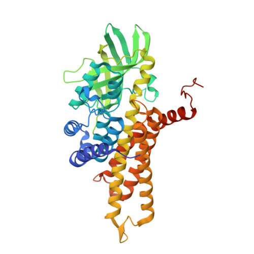
5AHS

3-Sulfinopropionyl-Coenzyme A (3SP-CoA) desulfinase from Advenella mimgardefordensis DPN7T: holo crystal structure with the substrate analog succinyl-CoA


Experimental Data Snapshot

  • Method: X-RAY DIFFRACTION
  • Resolution: 2.30 Å
  • R-Value Free: 0.244 
  • R-Value Work: 0.200 
  • R-Value Observed: 0.202 

Starting Model: experimental
View more details

wwPDB Validation   3D Report Full Report


Ligand Structure Quality Assessment 


This is version 1.4 of the entry. See complete history


Literature

3-Sulfinopropionyl-Coenzyme a (3Sp-Coa) Desulfinase from Advenella Mimigardefordensis Dpn7(T): Crystal Structure and Function of a Desulfinase with an Acyl-Coa Dehydrogenase Fold.

Schurmann, M.Meijers, R.Schneider, T.R.Steinbuchel, A.Cianci, M.

(2015) Acta Crystallogr D Biol Crystallogr 71: 1360

  • DOI: https://doi.org/10.1107/S1399004715006616
  • Primary Citation of Related Structures:  
    5AF7, 5AHS

  • PubMed Abstract: 

    3-Sulfinopropionyl-coenzyme A (3SP-CoA) desulfinase (AcdDPN7; EC 3.13.1.4) was identified during investigation of the 3,3'-dithiodipropionic acid (DTDP) catabolic pathway in the betaproteobacterium Advenella mimigardefordensis strain DPN7(T). DTDP is an organic disulfide and a precursor for the synthesis of polythioesters (PTEs) in bacteria, and is of interest for biotechnological PTE production. AcdDPN7 catalyzes sulfur abstraction from 3SP-CoA, a key step during the catabolism of DTDP. Here, the crystal structures of apo AcdDPN7 at 1.89 Å resolution and of its complex with the CoA moiety from the substrate analogue succinyl-CoA at 2.30 Å resolution are presented. The apo structure shows that AcdDPN7 belongs to the acyl-CoA dehydrogenase superfamily fold and that it is a tetramer, with each subunit containing one flavin adenine dinucleotide (FAD) molecule. The enzyme does not show any dehydrogenase activity. Dehydrogenase activity would require a catalytic base (Glu or Asp residue) at either position 246 or position 366, where a glutamine and a glycine are instead found, respectively, in this desulfinase. The positioning of CoA in the crystal complex enabled the modelling of a substrate complex containing 3SP-CoA. This indicates that Arg84 is a key residue in the desulfination reaction. An Arg84Lys mutant showed a complete loss of enzymatic activity, suggesting that the guanidinium group of the arginine is essential for desulfination. AcdDPN7 is the first desulfinase with an acyl-CoA dehydrogenase fold to be reported, which underlines the versatility of this enzyme scaffold.


  • Organizational Affiliation

    Institut für Molekulare Mikrobiologie und Biotechnologie, Westfälische Wilhelms-Universität Münster, 48149 Münster, Germany.


Macromolecules
Find similar proteins by:  (by identity cutoff)  |  3D Structure
Entity ID: 1
MoleculeChains Sequence LengthOrganismDetailsImage
ACYL-COA DEHYDROGENASE
A, B, C, D, E
A, B, C, D, E, F
401Advenella mimigardefordensis DPN7Mutation(s): 0 
EC: 3.13.1.4
UniProt
Find proteins for K4L7X3 (Advenella mimigardefordensis (strain DSM 17166 / LMG 22922 / DPN7))
Explore K4L7X3 
Go to UniProtKB:  K4L7X3
Entity Groups  
Sequence Clusters30% Identity50% Identity70% Identity90% Identity95% Identity100% Identity
UniProt GroupK4L7X3
Sequence Annotations
Expand
  • Reference Sequence
Small Molecules
Ligands 4 Unique
IDChains Name / Formula / InChI Key2D Diagram3D Interactions
FAD
Query on FAD

Download Ideal Coordinates CCD File 
G [auth A]
I [auth B]
K [auth C]
N [auth D]
P [auth E]
G [auth A],
I [auth B],
K [auth C],
N [auth D],
P [auth E],
R [auth F]
FLAVIN-ADENINE DINUCLEOTIDE
C27 H33 N9 O15 P2
VWWQXMAJTJZDQX-UYBVJOGSSA-N
COA
Query on COA

Download Ideal Coordinates CCD File 
H [auth A],
J [auth B],
O [auth D],
Q [auth E],
S [auth F]
COENZYME A
C21 H36 N7 O16 P3 S
RGJOEKWQDUBAIZ-IBOSZNHHSA-N
SIN
Query on SIN

Download Ideal Coordinates CCD File 
L [auth C]SUCCINIC ACID
C4 H6 O4
KDYFGRWQOYBRFD-UHFFFAOYSA-N
SO4
Query on SO4

Download Ideal Coordinates CCD File 
M [auth C]SULFATE ION
O4 S
QAOWNCQODCNURD-UHFFFAOYSA-L
Experimental Data & Validation

Experimental Data

  • Method: X-RAY DIFFRACTION
  • Resolution: 2.30 Å
  • R-Value Free: 0.244 
  • R-Value Work: 0.200 
  • R-Value Observed: 0.202 
  • Space Group: P 21 21 2
Unit Cell:
Length ( Å )Angle ( ˚ )
a = 100.066α = 90
b = 233.493β = 90
c = 121.285γ = 90
Software Package:
Software NamePurpose
PHENIXrefinement
XDSdata reduction
Aimlessdata scaling
PHASERphasing

Structure Validation

View Full Validation Report


Ligand Structure Quality Assessment 


Entry History 

Deposition Data

Revision History  (Full details and data files)

  • Version 1.0: 2015-06-03
    Type: Initial release
  • Version 1.1: 2015-06-17
    Changes: Database references
  • Version 1.2: 2017-08-23
    Changes: Data collection
  • Version 1.3: 2019-05-08
    Changes: Data collection, Experimental preparation
  • Version 1.4: 2024-01-10
    Changes: Data collection, Database references, Derived calculations, Other, Refinement description