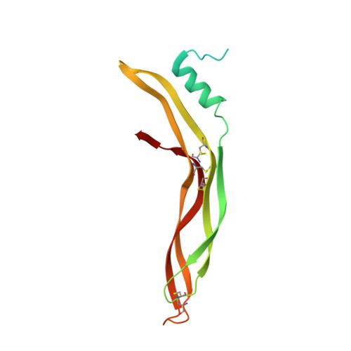
Structure of Gremlin-1 and Analysis of its Interaction with Bmp-2.
Kisonaite, M., Wang, X., Hyvonen, M.(2016) Biochem J 473: 1593
- PubMed: 27036124
- DOI: https://doi.org/10.1042/BCJ20160254
- Primary Citation of Related Structures:
5AEJ - PubMed Abstract:
Bone morphogenetic protein 2 (BMP-2) is a member of the transforming growth factor-β (TGF-β) signalling family and has a very broad biological role in development. Its signalling is regulated by many effectors: transmembrane proteins, membrane-attached proteins and soluble secreted antagonists such as Gremlin-1. Very little is known about the molecular mechanism by which Gremlin-1 and other DAN (differential screening-selected gene aberrative in neuroblastoma) family proteins inhibit BMP signalling. We analysed the interaction of Gremlin-1 with BMP-2 using a range of biophysical techniques, and used mutagenesis to map the binding site on BMP-2. We have also determined the crystal structure of Gremlin-1, revealing a similar conserved dimeric structure to that seen in other DAN family inhibitors. Measurements using biolayer interferometry (BLI) indicate that Gremlin-1 and BMP-2 can form larger complexes, beyond the expected 1:1 stoichiometry of dimers, forming oligomers that assemble in alternating fashion. These results suggest that inhibition of BMP-2 by Gremlin-1 occurs by a mechanism that is distinct from other known inhibitors such as Noggin and Chordin and we propose a novel model of BMP-2-Gremlin-1 interaction yet not seen among any BMP antagonists, and cannot rule out that several different oligomeric states could be found, depending on the concentration of the two proteins.
- Department of Biochemistry, University of Cambridge, 80 Tennis Court Road, Cambridge CB2 1GA, U.K.
Organizational Affiliation:


