5ADT | pdb_00005adt

Crystal structure of human tankyrase 2 in complex with OD73


Experimental Data Snapshot

  • Method: X-RAY DIFFRACTION
  • Resolution: 2.15 Å
  • R-Value Free: 
    0.217 (Depositor), 0.219 (DCC) 
  • R-Value Work: 
    0.173 (Depositor) 
  • R-Value Observed: 
    0.175 (Depositor) 

wwPDB Validation 3D Report Full Report

Validation slider image for 5ADT

Ligand Structure Quality Assessment 


This is version 1.2 of the entry. See complete history

Literature

Development and Structural Analysis of Adenosine Site Binding Tankyrase Inhibitors.

Haikarainen, T.Waaler, J.Ignatev, A.Nkizinkiko, Y.Venkannagari, H.Obaji, E.Krauss, S.Lehtio, L.

(2016) Bioorg Med Chem Lett 26: 328

  • DOI: https://doi.org/10.1016/j.bmcl.2015.12.018
  • Primary Citation Related Structures: 
    5ADQ, 5ADR, 5ADS, 5ADT, 5AEH

  • PubMed Abstract: 

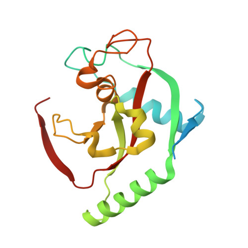
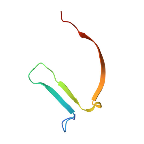
    Tankyrases 1 and 2, the specialized members of the ARTD protein family, are druggable biotargets whose inhibition may have therapeutic potential against cancer, metabolic disease, fibrotic disease, fibrotic wound healing and HSV viral infections. We have previously identified a novel tankyrase inhibitor scaffold, JW55, and showed that it reduces mouse colon adenoma formation in vivo. Here we expanded the scaffold and profiled the selectivity of the compounds against a panel of human ARTDs. The scaffold also enables a fine modulation of selectivity towards either tankyrase 1 or tankyrase 2. In order to get insight about the binding mode of the inhibitors, we solved crystal structures of the compounds in complex with tankyrase 2. The compounds bind to the adenosine pocket of the catalytic domain and cause changes in the protein structure that are modulated by the chemical modifications of the compounds. The structural analysis allows further rational development of this compound class as a potent and selective tankyrase inhibitor.


  • Organizational Affiliation
    • Faculty of Biochemistry and Molecular Medicine and Biocenter Oulu, University of Oulu, Oulu, Finland.

Macromolecule Content 

  • Total Structure Weight: 27.99 kDa 
  • Atom Count: 1,846 
  • Modeled Residue Count: 208 
  • Deposited Residue Count: 239 
  • Unique protein chains: 2

Macromolecules

Find similar proteins by:|  3D Structure
Entity ID: 1
MoleculeChains  Sequence LengthOrganismDetailsImage
TANKYRASE-2191Homo sapiensMutation(s): 0 
EC: 2.4.2 (UniProt), 2.4.2.30 (UniProt)
UniProt & NIH Common Fund Data Resources
Find proteins for Q9H2K2 (Homo sapiens)
Explore Q9H2K2 
Go to UniProtKB:  Q9H2K2
PHAROS:  Q9H2K2
GTEx:  ENSG00000107854 
Entity Groups
Sequence Clusters30% Identity50% Identity70% Identity90% Identity95% Identity100% Identity
UniProt GroupQ9H2K2
Sequence Annotations
Expand
Reference Sequence
Find similar proteins by:|  3D Structure
Entity ID: 2
MoleculeChains  Sequence LengthOrganismDetailsImage
TANKYRASE-248Homo sapiensMutation(s): 0 
EC: 2.4.2 (UniProt), 2.4.2.30 (UniProt)
UniProt & NIH Common Fund Data Resources
Find proteins for Q9H2K2 (Homo sapiens)
Explore Q9H2K2 
Go to UniProtKB:  Q9H2K2
PHAROS:  Q9H2K2
GTEx:  ENSG00000107854 
Entity Groups
Sequence Clusters30% Identity50% Identity70% Identity90% Identity95% Identity100% Identity
UniProt GroupQ9H2K2
Sequence Annotations
Expand
Reference Sequence

Small Molecules

Ligands 4 Unique
IDChains Name / Formula / InChI Key2D Diagram3D Interactions
1TC

Query on 1TC


Download:Ideal Coordinates CCD File
E [auth A]N-[3-chloranyl-4-[[4-(4-methoxyphenyl)oxan-4-yl]methylcarbamoyl]phenyl]-2-methyl-1,3-oxazole-5-carboxamide
C25 H26 Cl N3 O5
WOVOBLGUSQNZJX-UHFFFAOYSA-N
SO4

Query on SO4


Download:Ideal Coordinates CCD File
D [auth A],
G [auth B]
SULFATE ION
O4 S
QAOWNCQODCNURD-UHFFFAOYSA-L
ZN

Query on ZN


Download:Ideal Coordinates CCD File
F [auth A]ZINC ION
Zn
PTFCDOFLOPIGGS-UHFFFAOYSA-N
BCT

Query on BCT


Download:Ideal Coordinates CCD File
C [auth A]BICARBONATE ION
C H O3
BVKZGUZCCUSVTD-UHFFFAOYSA-M
Binding Affinity Annotations 
IDSourceBinding Affinity
1TC BindingDB:  5ADT IC50: min: 690, max: 2239 (nM) from 5 assay(s)

Experimental Data & Validation

Experimental Data

  • Method: X-RAY DIFFRACTION
  • Resolution: 2.15 Å
  • R-Value Free:  0.217 (Depositor), 0.219 (DCC) 
  • R-Value Work:  0.173 (Depositor) 
  • R-Value Observed: 0.175 (Depositor) 
Space Group: P 41 21 2
Unit Cell:
Length ( Å )Angle ( ˚ )
a = 66.33α = 90
b = 66.33β = 90
c = 120.84γ = 90
Software Package:
Software NamePurpose
REFMACrefinement
XDSdata reduction
XSCALEdata scaling

Structure Validation

View Full Validation Report


Ligand Structure Quality Assessment 


Entry History 

Deposition Data

Revision History  (Full details and data files)

  • Version 1.0: 2016-01-13
    Type: Initial release
  • Version 1.1: 2016-01-20
    Changes: Database references
  • Version 1.2: 2024-05-08
    Changes: Data collection, Database references, Derived calculations, Other