5ABJ | pdb_00005abj

Structure of Coxsackievirus A16 in complex with GPP3


Experimental Data Snapshot

  • Method: X-RAY DIFFRACTION
  • Resolution: 2.75 Å
  • R-Value Free: 
    0.307 (Depositor), 0.298 (DCC) 
  • R-Value Work: 
    0.305 (Depositor), 0.302 (DCC) 
  • R-Value Observed: 
    0.305 (Depositor) 

wwPDB Validation 3D Report Full Report

Validation slider image for 5ABJ

Ligand Structure Quality Assessment 


This is version 1.2 of the entry. See complete history

Literature

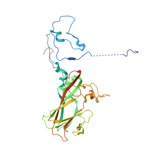
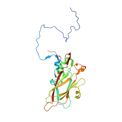
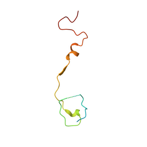
Structure Elucidation of Coxsackievirus A16 in Complex with Gpp3 Informs a Systematic Review of Highly Potent Capsid Binders to Enteroviruses.

De Colibus, L.Wang, X.Tijsma, A.Neyts, J.Spyrou, J.A.B.Ren, J.Grimes, J.M.Puerstinger, G.Leyssen, P.Fry, E.E.Rao, Z.Stuart, D.I.

(2015) PLoS Pathog 11: 5165

  • DOI: https://doi.org/10.1371/journal.ppat.1005165
  • Primary Citation Related Structures: 
    5ABJ

  • PubMed Abstract: 

    The replication of enterovirus 71 (EV71) and coxsackievirus A16 (CVA16), which are the major cause of hand, foot and mouth disease (HFMD) in children, can be inhibited by the capsid binder GPP3. Here, we present the crystal structure of CVA16 in complex with GPP3, which clarifies the role of the key residues involved in interactions with the inhibitor. Based on this model, in silico docking was performed to investigate the interactions with the two next-generation capsid binders NLD and ALD, which we show to be potent inhibitors of a panel of enteroviruses with potentially interesting pharmacological properties. A meta-analysis was performed using the available structural information to obtain a deeper insight into those structural features required for capsid binders to interact effectively and also those that confer broad-spectrum anti-enterovirus activity.


  • Organizational Affiliation
    • Division of Structural Biology, University of Oxford, Oxford, United Kingdom.

Macromolecule Content 

  • Total Structure Weight: 95.31 kDa 
  • Atom Count: 6,440 
  • Modeled Residue Count: 824 
  • Deposited Residue Count: 862 
  • Unique protein chains: 4

Macromolecules

Find similar proteins by:|  3D Structure
Entity ID: 1
MoleculeChains  Sequence LengthOrganismDetailsImage
VP1297Coxsackievirus A16Mutation(s): 0 
UniProt
Find proteins for I3W9E1 (Coxsackievirus A16)
Explore I3W9E1 
Go to UniProtKB:  I3W9E1
Entity Groups
Sequence Clusters30% Identity50% Identity70% Identity90% Identity95% Identity100% Identity
UniProt GroupI3W9E1
Sequence Annotations
Expand
Reference Sequence
Find similar proteins by:|  3D Structure
Entity ID: 2
MoleculeChains  Sequence LengthOrganismDetailsImage
VP2254Coxsackievirus A16Mutation(s): 0 
UniProt
Find proteins for I3W9E1 (Coxsackievirus A16)
Explore I3W9E1 
Go to UniProtKB:  I3W9E1
Entity Groups
Sequence Clusters30% Identity50% Identity70% Identity90% Identity95% Identity100% Identity
UniProt GroupI3W9E1
Sequence Annotations
Expand
Reference Sequence
Find similar proteins by:|  3D Structure
Entity ID: 3
MoleculeChains  Sequence LengthOrganismDetailsImage
VP3242Coxsackievirus A16Mutation(s): 0 
UniProt
Find proteins for I3W9E1 (Coxsackievirus A16)
Explore I3W9E1 
Go to UniProtKB:  I3W9E1
Entity Groups
Sequence Clusters30% Identity50% Identity70% Identity90% Identity95% Identity100% Identity
UniProt GroupI3W9E1
Sequence Annotations
Expand
Reference Sequence
Find similar proteins by:|  3D Structure
Entity ID: 4
MoleculeChains  Sequence LengthOrganismDetailsImage
VP469Coxsackievirus A16Mutation(s): 0 
UniProt
Find proteins for I3W9E1 (Coxsackievirus A16)
Explore I3W9E1 
Go to UniProtKB:  I3W9E1
Entity Groups
Sequence Clusters30% Identity50% Identity70% Identity90% Identity95% Identity100% Identity
UniProt GroupI3W9E1
Sequence Annotations
Expand
Reference Sequence

Small Molecules

Experimental Data & Validation

Experimental Data

  • Method: X-RAY DIFFRACTION
  • Resolution: 2.75 Å
  • R-Value Free:  0.307 (Depositor), 0.298 (DCC) 
  • R-Value Work:  0.305 (Depositor), 0.302 (DCC) 
  • R-Value Observed: 0.305 (Depositor) 
Space Group: P 41 21 2
Unit Cell:
Length ( Å )Angle ( ˚ )
a = 491.6α = 90
b = 491.6β = 90
c = 709.56γ = 90
Software Package:
Software NamePurpose
CNSrefinement
HKL-2000data reduction
SCALEPACKdata scaling
CNSphasing

Structure Validation

View Full Validation Report


Ligand Structure Quality Assessment 


Entry History 

Deposition Data

Revision History  (Full details and data files)

  • Version 1.0: 2015-09-09
    Type: Initial release
  • Version 1.1: 2015-11-04
    Changes: Database references
  • Version 1.2: 2024-05-08
    Changes: Data collection, Database references, Derived calculations, Other