5AA2

Crystal structure of MltF from Pseudomonas aeruginosa in complex with NAM-pentapeptide.


Experimental Data Snapshot

  • Method: X-RAY DIFFRACTION
  • Resolution: 2.80 Å
  • R-Value Free: 0.235 
  • R-Value Work: 0.167 
  • R-Value Observed: 0.171 

wwPDB Validation   3D Report Full Report


This is version 2.1 of the entry. See complete history


Literature

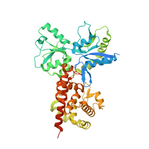
Activation by Allostery in Cell-Wall Remodeling by a Modular Membrane-Bound Lytic Transglycosylase from Pseudomonas aeruginosa.

Dominguez-Gil, T.Lee, M.Acebron-Avalos, I.Mahasenan, K.V.Hesek, D.Dik, D.A.Byun, B.Lastochkin, E.Fisher, J.F.Mobashery, S.Hermoso, J.A.

(2016) Structure 24: 1729-1741

  • DOI: https://doi.org/10.1016/j.str.2016.07.019
  • Primary Citation of Related Structures:  
    5A5X, 5AA1, 5AA2, 5AA3, 5AA4

  • PubMed Abstract: 

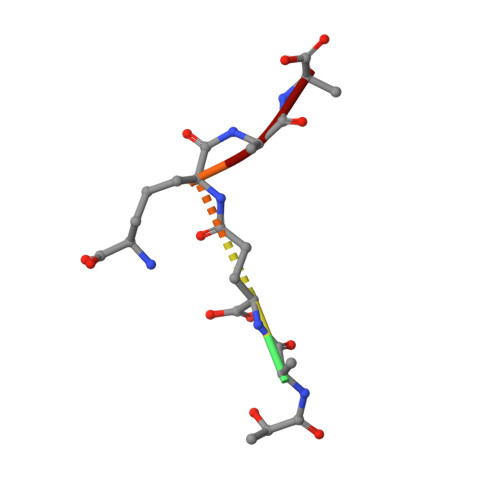
    Bacteria grow and divide without loss of cellular integrity. This accomplishment is notable, as a key component of their cell envelope is a surrounding glycopeptide polymer. In Gram-negative bacteria this polymer-the peptidoglycan-grows by the difference between concurrent synthesis and degradation. The regulation of the enzymatic ensemble for these activities is poorly understood. We report herein the structural basis for the control of one such enzyme, the lytic transglycosylase MltF of Pseudomonas aeruginosa. Its structure comprises two modules: an ABC-transporter-like regulatory module and a catalytic module. Occupancy of the regulatory module by peptidoglycan-derived muropeptides effects a dramatic and long-distance (40 Å) conformational change, occurring over the entire protein structure, to open its active site for catalysis. This discovery of the molecular basis for the allosteric control of MltF catalysis is foundational to further study of MltF within the complex enzymatic orchestration of the dynamic peptidoglycan.


  • Organizational Affiliation

    Departamento de Cristalografía y Biología Estructural, Instituto de Química-Física "Rocasolano", CSIC, Serrano 119, 28006 Madrid, Spain.


Macromolecules
Find similar proteins by:  (by identity cutoff)  |  3D Structure
Entity ID: 1
MoleculeChains Sequence LengthOrganismDetailsImage
MEMBRANE-BOUND LYTIC MUREIN TRANSGLYCOSYLASE F
A, C
499Pseudomonas aeruginosa BWHPSA013Mutation(s): 0 
EC: 4.2.2
UniProt
Find proteins for Q9HXN1 (Pseudomonas aeruginosa (strain ATCC 15692 / DSM 22644 / CIP 104116 / JCM 14847 / LMG 12228 / 1C / PRS 101 / PAO1))
Explore Q9HXN1 
Go to UniProtKB:  Q9HXN1
Entity Groups  
Sequence Clusters30% Identity50% Identity70% Identity90% Identity95% Identity100% Identity
UniProt GroupQ9HXN1
Sequence Annotations
Expand
  • Reference Sequence
Find similar proteins by:  (by identity cutoff)  |  3D Structure
Entity ID: 2
MoleculeChains Sequence LengthOrganismDetailsImage
MEMBRANE-BOUND LYTIC MUREIN TRANSGLYCOSYLASE F499Pseudomonas aeruginosa BWHPSA013Mutation(s): 0 
EC: 4.2.2
UniProt
Find proteins for Q9HXN1 (Pseudomonas aeruginosa (strain ATCC 15692 / DSM 22644 / CIP 104116 / JCM 14847 / LMG 12228 / 1C / PRS 101 / PAO1))
Explore Q9HXN1 
Go to UniProtKB:  Q9HXN1
Entity Groups  
Sequence Clusters30% Identity50% Identity70% Identity90% Identity95% Identity100% Identity
UniProt GroupQ9HXN1
Sequence Annotations
Expand
  • Reference Sequence
Find similar proteins by:  (by identity cutoff)  |  3D Structure
Entity ID: 3
MoleculeChains Sequence LengthOrganismDetailsImage
MEMBRANE-BOUND LYTIC MUREIN TRANSGLYCOSYLASE F499Pseudomonas aeruginosa BWHPSA013Mutation(s): 0 
EC: 4.2.2
UniProt
Find proteins for Q9HXN1 (Pseudomonas aeruginosa (strain ATCC 15692 / DSM 22644 / CIP 104116 / JCM 14847 / LMG 12228 / 1C / PRS 101 / PAO1))
Explore Q9HXN1 
Go to UniProtKB:  Q9HXN1
Entity Groups  
Sequence Clusters30% Identity50% Identity70% Identity90% Identity95% Identity100% Identity
UniProt GroupQ9HXN1
Sequence Annotations
Expand
  • Reference Sequence

Find similar proteins by:  Sequence   |   3D Structure  

Entity ID: 4
MoleculeChains Sequence LengthOrganismDetailsImage
N-ACETYLGLUCOSAMINE-1,6-ANHYDRO-N-ACETYLMURAMIC ACID L-ALA-D-GLU-M-DAP-D-ALA-D-ALA6Pseudomonas aeruginosa BWHPSA013Mutation(s): 0 
Sequence Annotations
Expand
  • Reference Sequence
Small Molecules
Modified Residues  1 Unique
IDChains TypeFormula2D DiagramParent
API
Query on API
E
L-PEPTIDE LINKINGC7 H14 N2 O4LYS
Experimental Data & Validation

Experimental Data

  • Method: X-RAY DIFFRACTION
  • Resolution: 2.80 Å
  • R-Value Free: 0.235 
  • R-Value Work: 0.167 
  • R-Value Observed: 0.171 
  • Space Group: P 1 21 1
Unit Cell:
Length ( Å )Angle ( ˚ )
a = 65.585α = 90
b = 135.652β = 92.06
c = 137.459γ = 90
Software Package:
Software NamePurpose
PHENIXrefinement
XDSdata reduction
Aimlessdata scaling
PHASERphasing

Structure Validation

View Full Validation Report


Entry History 

Deposition Data

Revision History  (Full details and data files)

  • Version 1.0: 2016-10-12
    Type: Initial release
  • Version 1.1: 2017-05-03
    Changes: Database references
  • Version 2.0: 2023-11-15
    Changes: Atomic model, Data collection, Database references, Derived calculations, Other
  • Version 2.1: 2024-01-10
    Changes: Refinement description