4ZYP | pdb_00004zyp

Crystal Structure of Motavizumab and Quaternary-Specific RSV-Neutralizing Human Antibody AM14 in Complex with Prefusion RSV F Glycoprotein


Experimental Data Snapshot

  • Method: X-RAY DIFFRACTION
  • Resolution: 5.50 Å
  • R-Value Free: 
    0.277 (Depositor), 0.275 (DCC) 
  • R-Value Work: 
    0.210 (Depositor), 0.215 (DCC) 
  • R-Value Observed: 
    0.214 (Depositor) 

Starting Models: experimental
View more details

wwPDB Validation 3D Report Full Report

Validation slider image for 4ZYP

This is version 1.2 of the entry. See complete history

Literature

Characterization of a Prefusion-Specific Antibody That Recognizes a Quaternary, Cleavage-Dependent Epitope on the RSV Fusion Glycoprotein.

Gilman, M.S.Moin, S.M.Mas, V.Chen, M.Patel, N.K.Kramer, K.Zhu, Q.Kabeche, S.C.Kumar, A.Palomo, C.Beaumont, T.Baxa, U.Ulbrandt, N.D.Melero, J.A.Graham, B.S.McLellan, J.S.

(2015) PLoS Pathog 11: e1005035-e1005035

  • DOI: https://doi.org/10.1371/journal.ppat.1005035
  • Primary Citation Related Structures: 
    4ZYK, 4ZYP

  • PubMed Abstract: 

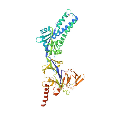
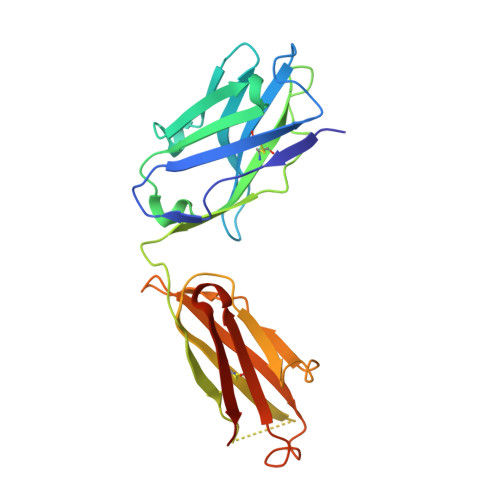
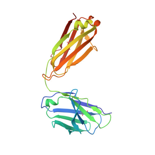
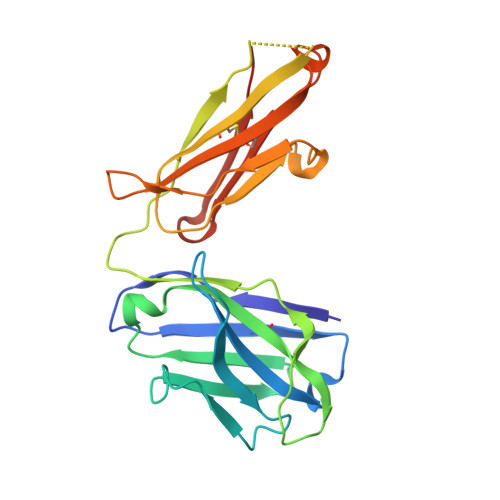
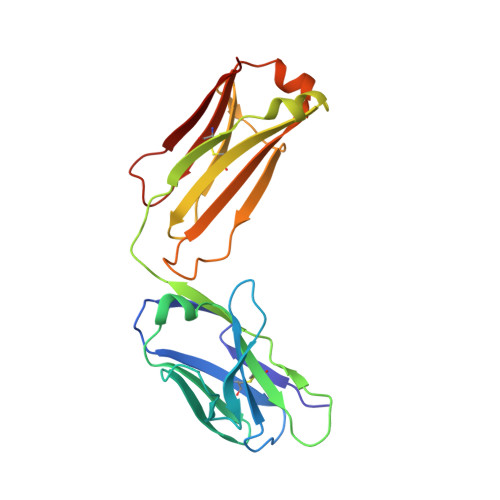
    Prevention efforts for respiratory syncytial virus (RSV) have been advanced due to the recent isolation and characterization of antibodies that specifically recognize the prefusion conformation of the RSV fusion (F) glycoprotein. These potently neutralizing antibodies are in clinical development for passive prophylaxis and have also aided the design of vaccine antigens that display prefusion-specific epitopes. To date, prefusion-specific antibodies have been shown to target two antigenic sites on RSV F, but both of these sites are also present on monomeric forms of F. Here we present a structural and functional characterization of human antibody AM14, which potently neutralized laboratory strains and clinical isolates of RSV from both A and B subtypes. The crystal structure and location of escape mutations revealed that AM14 recognizes a quaternary epitope that spans two protomers and includes a region that undergoes extensive conformational changes in the pre- to postfusion F transition. Binding assays demonstrated that AM14 is unique in its specific recognition of trimeric furin-cleaved prefusion F, which is the mature form of F on infectious virions. These results demonstrate that the prefusion F trimer contains potent neutralizing epitopes not present on monomers and that AM14 should be particularly useful for characterizing the conformational state of RSV F-based vaccine antigens.


  • Organizational Affiliation
    • Department of Biochemistry, Geisel School of Medicine at Dartmouth, Hanover, New Hampshire, United States of America.

Macromolecule Content 

  • Total Structure Weight: 451.61 kDa 
  • Atom Count: 29,990 
  • Modeled Residue Count: 3,906 
  • Deposited Residue Count: 4,134 
  • Unique protein chains: 5

Macromolecules

Find similar proteins by:|  3D Structure
Entity ID: 1
MoleculeChains  Sequence LengthOrganismDetailsImage
Fusion glycoprotein F0,Fibritin
A, B, C
498Human respiratory syncytial virus A2Tequatrovirus T4
This entity is chimeric
Mutation(s): 6 
UniProt
Find proteins for D9IEJ2 (Enterobacteria phage T4)
Explore D9IEJ2 
Go to UniProtKB:  D9IEJ2
Find proteins for P03420 (Human respiratory syncytial virus A (strain A2))
Explore P03420 
Go to UniProtKB:  P03420
Entity Groups
Sequence Clusters30% Identity50% Identity70% Identity90% Identity95% Identity100% Identity
UniProt GroupsP03420D9IEJ2
Sequence Annotations
Expand
Reference Sequence
Find similar proteins by:|  3D Structure
Entity ID: 2
MoleculeChains  Sequence LengthOrganismDetailsImage
Motavizumab antibody Fab heavy chainD [auth J],
L [auth K],
N
225Mus musculusMutation(s): 0 
Find similar proteins by:|  3D Structure
Entity ID: 3
MoleculeChains  Sequence LengthOrganismDetailsImage
Motavizumab antibody light chainE [auth L],
M,
O
213Mus musculusMutation(s): 0 
Find similar proteins by:|  3D Structure
Entity ID: 4
MoleculeChains  Sequence LengthOrganismDetailsImage
AM14 antibody Fab heavy chainF,
H,
J [auth D]
227Homo sapiensMutation(s): 0 
Find similar proteins by:|  3D Structure
Entity ID: 5
MoleculeChains  Sequence LengthOrganismDetailsImage
AM14 antibody light chainG,
I,
K [auth E]
215Homo sapiensMutation(s): 0 

Experimental Data & Validation

Experimental Data

  • Method: X-RAY DIFFRACTION
  • Resolution: 5.50 Å
  • R-Value Free:  0.277 (Depositor), 0.275 (DCC) 
  • R-Value Work:  0.210 (Depositor), 0.215 (DCC) 
  • R-Value Observed: 0.214 (Depositor) 
Space Group: P 1 21 1
Unit Cell:
Length ( Å )Angle ( ˚ )
a = 114.38α = 90
b = 210.29β = 100.46
c = 118.2γ = 90
Software Package:
Software NamePurpose
PHENIXrefinement
Aimlessdata scaling
PDB_EXTRACTdata extraction
Cootmodel building
PHASERphasing
MOSFLMdata reduction

Structure Validation

View Full Validation Report


Entry History 

Deposition Data

Revision History  (Full details and data files)

  • Version 1.0: 2015-07-29
    Type: Initial release
  • Version 1.1: 2023-09-27
    Changes: Data collection, Database references, Derived calculations, Refinement description, Source and taxonomy
  • Version 1.2: 2024-11-20
    Changes: Structure summary