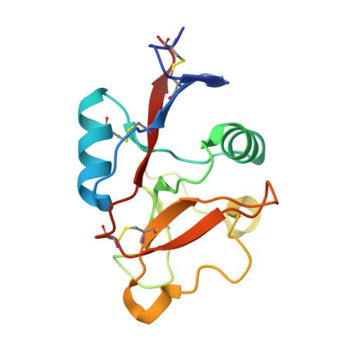
4ZRV

Structure of cow mincle CRD complexed with trehalose mono butyrate


Experimental Data Snapshot

  • Method: X-RAY DIFFRACTION
  • Resolution: 2.10 Å
  • R-Value Free: 0.208 
  • R-Value Work: 0.163 
  • R-Value Observed: 0.166 

wwPDB Validation   3D Report Full Report


This is version 2.2 of the entry. See complete history


Literature

Binding Sites for Acylated Trehalose Analogs of Glycolipid Ligands on an Extended Carbohydrate Recognition Domain of the Macrophage Receptor Mincle.

Feinberg, H.Rambaruth, N.D.Jegouzo, S.A.Jacobsen, K.M.Djurhuus, R.Poulsen, T.B.Weis, W.I.Taylor, M.E.Drickamer, K.

(2016) J Biol Chem 291: 21222-21233

  • DOI: https://doi.org/10.1074/jbc.M116.749515
  • Primary Citation of Related Structures:  
    4ZRV, 4ZRW, 5KTH, 5KTI

  • PubMed Abstract: 

    The macrophage receptor mincle binds to trehalose dimycolate on the surface of Mycobacterium tuberculosis Signaling initiated by this interaction leads to cytokine production, which underlies the ability of mycobacteria to evade the immune system and also to function as adjuvants. In previous work the mechanism for binding of the sugar headgroup of trehalose dimycolate to mincle has been elucidated, but the basis for enhanced binding to glycolipid ligands, in which hydrophobic substituents are attached to the 6-hydroxyl groups, has been the subject of speculation. In the work reported here, the interaction of trehalose derivatives with bovine mincle has been probed with a series of synthetic mimics of trehalose dimycolate in binding assays, in structural studies by x-ray crystallography, and by site-directed mutagenesis. Binding studies reveal that, rather than reflecting specific structural preference, the apparent affinity of mincle for ligands with hydrophobic substituents correlates with their overall size. Structural and mutagenesis analysis provides evidence for interaction of the hydrophobic substituents with multiple different portions of the surface of mincle and confirms the presence of three Ca 2+ -binding sites. The structure of an extended portion of the extracellular domain of mincle, beyond the minimal C-type carbohydrate recognition domain, also constrains the way the binding domains may interact on the surface of macrophages.


  • Organizational Affiliation

    From the Departments of Structural Biology and Molecular and Cellular Physiology, Stanford University School of Medicine, Stanford, California 94305.


Macromolecules
Find similar proteins by:  (by identity cutoff)  |  3D Structure
Entity ID: 1
MoleculeChains Sequence LengthOrganismDetailsImage
Mincle CRD
A, B, C
134Bos taurusMutation(s): 1 
Gene Names: CLEC4E
UniProt
Find proteins for E1BHM0 (Bos taurus)
Explore E1BHM0 
Go to UniProtKB:  E1BHM0
Entity Groups  
Sequence Clusters30% Identity50% Identity70% Identity90% Identity95% Identity100% Identity
UniProt GroupE1BHM0
Sequence Annotations
Expand
  • Reference Sequence
Oligosaccharides

Help

Entity ID: 2
MoleculeChains Length2D Diagram Glycosylation3D Interactions
6-O-butanoyl-alpha-D-glucopyranose-(1-1)-alpha-D-glucopyranose
D, E, F
2N/AN/A
Glycosylation Resources
GlyTouCan:  G42831FS
GlyCosmos:  G42831FS
Small Molecules
Ligands 2 Unique
IDChains Name / Formula / InChI Key2D Diagram3D Interactions
ACT
Query on ACT

Download Ideal Coordinates CCD File 
J [auth A],
N [auth C],
R [auth C]
ACETATE ION
C2 H3 O2
QTBSBXVTEAMEQO-UHFFFAOYSA-M
CA
Query on CA

Download Ideal Coordinates CCD File 
G [auth A]
H [auth A]
I [auth A]
K [auth B]
L [auth B]
G [auth A],
H [auth A],
I [auth A],
K [auth B],
L [auth B],
M [auth B],
O [auth C],
P [auth C],
Q [auth C]
CALCIUM ION
Ca
BHPQYMZQTOCNFJ-UHFFFAOYSA-N
Biologically Interesting Molecules (External Reference) 1 Unique
Experimental Data & Validation

Experimental Data

  • Method: X-RAY DIFFRACTION
  • Resolution: 2.10 Å
  • R-Value Free: 0.208 
  • R-Value Work: 0.163 
  • R-Value Observed: 0.166 
  • Space Group: P 1 21 1
Unit Cell:
Length ( Å )Angle ( ˚ )
a = 66.464α = 90
b = 46.676β = 101.16
c = 77.92γ = 90
Software Package:
Software NamePurpose
SCALAdata scaling
PHENIXrefinement
PDB_EXTRACTdata extraction
XDSdata reduction

Structure Validation

View Full Validation Report


Entry History & Funding Information

Deposition Data


Funding OrganizationLocationGrant Number
Wellcome TrustUnited Kingdom093599

Revision History  (Full details and data files)

  • Version 1.0: 2016-05-25
    Type: Initial release
  • Version 1.1: 2016-08-31
    Changes: Database references
  • Version 1.2: 2016-10-19
    Changes: Database references
  • Version 2.0: 2020-07-29
    Type: Remediation
    Reason: Carbohydrate remediation
    Changes: Advisory, Atomic model, Data collection, Database references, Derived calculations, Non-polymer description, Structure summary
  • Version 2.1: 2023-08-09
    Changes: Data collection, Database references, Structure summary
  • Version 2.2: 2024-10-30
    Changes: Data collection, Structure summary