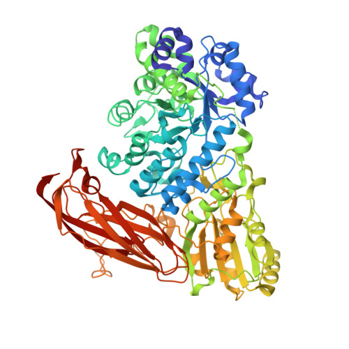
4ZOD

Crystal Structure of beta-glucosidase from Listeria innocua in complex with glucose


Experimental Data Snapshot

  • Method: X-RAY DIFFRACTION
  • Resolution: 2.10 Å
  • R-Value Free: 0.233 
  • R-Value Work: 0.167 
  • R-Value Observed: 0.170 

Starting Model: experimental
View more details

wwPDB Validation   3D Report Full Report


Ligand Structure Quality Assessment 


This is version 1.4 of the entry. See complete history


Literature

Functional and Structural Analysis of a beta-Glucosidase Involved in beta-1,2-Glucan Metabolism in Listeria innocua

Nakajima, M.Yoshida, R.Miyanaga, A.Abe, K.Takahashi, Y.Sugimoto, N.Toyoizumi, H.Nakai, H.Kitaoka, M.Taguchi, H.

(2016) PLoS One 11: e0148870-e0148870

  • DOI: https://doi.org/10.1371/journal.pone.0148870
  • Primary Citation of Related Structures:  
    4ZO6, 4ZO7, 4ZO8, 4ZO9, 4ZOA, 4ZOB, 4ZOC, 4ZOD, 4ZOE

  • PubMed Abstract: 

    Despite the presence of β-1,2-glucan in nature, few β-1,2-glucan degrading enzymes have been reported to date. Recently, the Lin1839 protein from Listeria innocua was identified as a 1,2-β-oligoglucan phosphorylase. Since the adjacent lin1840 gene in the gene cluster encodes a putative glycoside hydrolase family 3 β-glucosidase, we hypothesized that Lin1840 is also involved in β-1,2-glucan dissimilation. Here we report the functional and structural analysis of Lin1840. A recombinant Lin1840 protein (Lin1840r) showed the highest hydrolytic activity toward sophorose (Glc-β-1,2-Glc) among β-1,2-glucooligosaccharides, suggesting that Lin1840 is a β-glucosidase involved in sophorose degradation. The enzyme also rapidly hydrolyzed laminaribiose (β-1,3), but not cellobiose (β-1,4) or gentiobiose (β-1,6) among β-linked gluco-disaccharides. We determined the crystal structures of Lin1840r in complexes with sophorose and laminaribiose as productive binding forms. In these structures, Arg572 forms many hydrogen bonds with sophorose and laminaribiose at subsite +1, which seems to be a key factor for substrate selectivity. The opposite side of subsite +1 from Arg572 is connected to a large empty space appearing to be subsite +2 for the binding of sophorotriose (Glc-β-1,2-Glc-β-1,2-Glc) in spite of the higher Km value for sophorotriose than that for sophorose. The conformations of sophorose and laminaribiose are almost the same on the Arg572 side but differ on the subsite +2 side that provides no interaction with a substrate. Therefore, Lin1840r is unable to distinguish between sophorose and laminaribiose as substrates. These results provide the first mechanistic insights into β-1,2-glucooligosaccharide recognition by β-glucosidase.


  • Organizational Affiliation

    Department of Applied Biological Science, Faculty of Science and Technology, Tokyo University of Science, Noda, Chiba, Japan.


Macromolecules
Find similar proteins by:  (by identity cutoff)  |  3D Structure
Entity ID: 1
MoleculeChains Sequence LengthOrganismDetailsImage
Lin1840 protein
A, B
731Listeria innocua Clip11262Mutation(s): 1 
Gene Names: lin1840
EC: 3.2.1.21
UniProt
Find proteins for Q92AS9 (Listeria innocua serovar 6a (strain ATCC BAA-680 / CLIP 11262))
Explore Q92AS9 
Go to UniProtKB:  Q92AS9
Entity Groups  
Sequence Clusters30% Identity50% Identity70% Identity90% Identity95% Identity100% Identity
UniProt GroupQ92AS9
Sequence Annotations
Expand
  • Reference Sequence
Small Molecules
Ligands 5 Unique
IDChains Name / Formula / InChI Key2D Diagram3D Interactions
BGC
Query on BGC

Download Ideal Coordinates CCD File 
C [auth A],
L [auth B],
M [auth B]
beta-D-glucopyranose
C6 H12 O6
WQZGKKKJIJFFOK-VFUOTHLCSA-N
PEG
Query on PEG

Download Ideal Coordinates CCD File 
H [auth A],
K [auth B]
DI(HYDROXYETHYL)ETHER
C4 H10 O3
MTHSVFCYNBDYFN-UHFFFAOYSA-N
SO4
Query on SO4

Download Ideal Coordinates CCD File 
I [auth A]SULFATE ION
O4 S
QAOWNCQODCNURD-UHFFFAOYSA-L
GOL
Query on GOL

Download Ideal Coordinates CCD File 
E [auth A]
F [auth A]
G [auth A]
J [auth A]
O [auth B]
E [auth A],
F [auth A],
G [auth A],
J [auth A],
O [auth B],
P [auth B],
Q [auth B]
GLYCEROL
C3 H8 O3
PEDCQBHIVMGVHV-UHFFFAOYSA-N
MG
Query on MG

Download Ideal Coordinates CCD File 
D [auth A],
N [auth B]
MAGNESIUM ION
Mg
JLVVSXFLKOJNIY-UHFFFAOYSA-N
Experimental Data & Validation

Experimental Data

  • Method: X-RAY DIFFRACTION
  • Resolution: 2.10 Å
  • R-Value Free: 0.233 
  • R-Value Work: 0.167 
  • R-Value Observed: 0.170 
  • Space Group: I 1 2 1
Unit Cell:
Length ( Å )Angle ( ˚ )
a = 89.79α = 90
b = 95.41β = 97.27
c = 213.233γ = 90
Software Package:
Software NamePurpose
MOLREPphasing
REFMACrefinement
PDB_EXTRACTdata extraction

Structure Validation

View Full Validation Report


Ligand Structure Quality Assessment 


Entry History 

Deposition Data

Revision History  (Full details and data files)

  • Version 1.0: 2016-05-18
    Type: Initial release
  • Version 1.1: 2016-06-01
    Changes: Database references
  • Version 1.2: 2020-02-19
    Changes: Data collection, Derived calculations
  • Version 1.3: 2020-07-29
    Type: Remediation
    Reason: Carbohydrate remediation
    Changes: Data collection, Derived calculations, Structure summary
  • Version 1.4: 2023-11-08
    Changes: Data collection, Database references, Refinement description, Structure summary