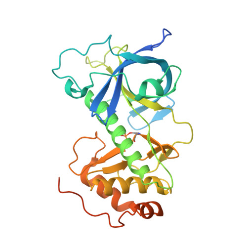
4ZME

Crystal Structure of the Alpha-kinase Domain of Myosin-II Heavy Chain Kinase A in Complex with Adenosine


Experimental Data Snapshot

  • Method: X-RAY DIFFRACTION
  • Resolution: 1.98 Å
  • R-Value Free: 0.236 
  • R-Value Work: 0.214 
  • R-Value Observed: 0.215 

Starting Model: experimental
View more details

wwPDB Validation   3D Report Full Report


Ligand Structure Quality Assessment 


This is version 1.4 of the entry. See complete history


Literature

Characterization of the Catalytic and Nucleotide Binding Properties of the alpha-Kinase Domain of Dictyostelium Myosin-II Heavy Chain Kinase A.

Yang, Y.Ye, Q.Jia, Z.Cote, G.P.

(2015) J Biol Chem 290: 23935-23946

  • DOI: https://doi.org/10.1074/jbc.M115.672410
  • Primary Citation of Related Structures:  
    4ZME, 4ZMF, 4ZS4

  • PubMed Abstract: 

    The α-kinases are a widely expressed family of serine/threonine protein kinases that exhibit no sequence identity with conventional eukaryotic protein kinases. In this report, we provide new information on the catalytic properties of the α-kinase domain of Dictyostelium myosin-II heavy chain kinase-A (termed A-CAT). Crystallization of A-CAT in the presence of MgATP yielded structures with AMP or adenosine in the catalytic cleft together with a phosphorylated Asp-766 residue. The results show that the β- and α-phosphoryl groups are transferred either directly or indirectly to the catalytically essential Asp-766. Biochemical assays confirmed that A-CAT hydrolyzed ATP, ADP, and AMP with kcat values of 1.9, 0.6, and 0.32 min(-1), respectively, and showed that A-CAT can use ADP to phosphorylate peptides and proteins. Binding assays using fluorescent 2'/3'-O-(N-methylanthraniloyl) analogs of ATP and ADP yielded Kd values for ATP, ADP, AMP, and adenosine of 20 ± 3, 60 ± 20, 160 ± 60, and 45 ± 15 μM, respectively. Site-directed mutagenesis showed that Glu-713, Leu-716, and Lys-645, all of which interact with the adenine base, were critical for nucleotide binding. Mutation of the highly conserved Gln-758, which chelates a nucleotide-associated Mg(2+) ion, eliminated catalytic activity, whereas loss of the highly conserved Lys-722 and Arg-592 decreased kcat values for kinase and ATPase activities by 3-6-fold. Mutation of Asp-663 impaired kinase activity to a much greater extent than ATPase, indicating a specific role in peptide substrate binding, whereas mutation of Gln-768 doubled ATPase activity, suggesting that it may act to exclude water from the active site.


  • Organizational Affiliation

    From the Department of Biomedical and Molecular Sciences, Queen's University, Kingston, Ontario K7L 3N6, Canada.


Macromolecules
Find similar proteins by:  (by identity cutoff)  |  3D Structure
Entity ID: 1
MoleculeChains Sequence LengthOrganismDetailsImage
Myosin heavy chain kinase A
A, B
307Dictyostelium discoideumMutation(s): 0 
Gene Names: mhkAmhckADDB_G0291231
EC: 2.7.11.7
UniProt
Find proteins for P42527 (Dictyostelium discoideum)
Explore P42527 
Go to UniProtKB:  P42527
Entity Groups  
Sequence Clusters30% Identity50% Identity70% Identity90% Identity95% Identity100% Identity
UniProt GroupP42527
Sequence Annotations
Expand
  • Reference Sequence
Small Molecules
Ligands 4 Unique
IDChains Name / Formula / InChI Key2D Diagram3D Interactions
ADN
Query on ADN

Download Ideal Coordinates CCD File 
D [auth A],
H [auth B]
ADENOSINE
C10 H13 N5 O4
OIRDTQYFTABQOQ-KQYNXXCUSA-N
PGE
Query on PGE

Download Ideal Coordinates CCD File 
K [auth B]TRIETHYLENE GLYCOL
C6 H14 O4
ZIBGPFATKBEMQZ-UHFFFAOYSA-N
PO4
Query on PO4

Download Ideal Coordinates CCD File 
E [auth A],
F [auth A],
I [auth B],
J [auth B]
PHOSPHATE ION
O4 P
NBIIXXVUZAFLBC-UHFFFAOYSA-K
ZN
Query on ZN

Download Ideal Coordinates CCD File 
C [auth A],
G [auth B]
ZINC ION
Zn
PTFCDOFLOPIGGS-UHFFFAOYSA-N
Modified Residues  1 Unique
IDChains TypeFormula2D DiagramParent
PHD
Query on PHD
A, B
L-PEPTIDE LINKINGC4 H8 N O7 PASP
Experimental Data & Validation

Experimental Data

  • Method: X-RAY DIFFRACTION
  • Resolution: 1.98 Å
  • R-Value Free: 0.236 
  • R-Value Work: 0.214 
  • R-Value Observed: 0.215 
  • Space Group: P 21 21 2
Unit Cell:
Length ( Å )Angle ( ˚ )
a = 83.194α = 90
b = 111.288β = 90
c = 79.56γ = 90
Software Package:
Software NamePurpose
REFMACrefinement
PDB_EXTRACTdata extraction
HKL-2000data reduction
HKL-2000data scaling
PHASERphasing

Structure Validation

View Full Validation Report


Ligand Structure Quality Assessment 


Entry History & Funding Information

Deposition Data

  • Released Date: 2015-08-19 
  • Deposition Author(s): Ye, Q., Jia, Z.

Funding OrganizationLocationGrant Number
Canadian Institutes of Health Research (CIHR)Canada--

Revision History  (Full details and data files)

  • Version 1.0: 2015-08-19
    Type: Initial release
  • Version 1.1: 2015-09-02
    Changes: Database references
  • Version 1.2: 2015-10-07
    Changes: Data collection, Database references
  • Version 1.3: 2020-01-08
    Changes: Author supporting evidence, Derived calculations
  • Version 1.4: 2023-09-27
    Changes: Data collection, Database references, Derived calculations, Refinement description