4ZHC

Siderocalin-mediated recognition and cellular uptake of actinides


Experimental Data Snapshot

  • Method: X-RAY DIFFRACTION
  • Resolution: 2.04 Å
  • R-Value Free: 0.229 
  • R-Value Work: 0.206 
  • R-Value Observed: 0.207 

wwPDB Validation   3D Report Full Report


Ligand Structure Quality Assessment 


This is version 1.3 of the entry. See complete history


Literature

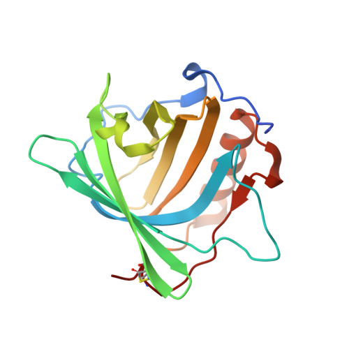
Siderocalin-mediated recognition, sensitization, and cellular uptake of actinides.

Allred, B.E.Rupert, P.B.Gauny, S.S.An, D.D.Ralston, C.Y.Sturzbecher-Hoehne, M.Strong, R.K.Abergel, R.J.

(2015) Proc Natl Acad Sci U S A 112: 10342-10347

  • DOI: https://doi.org/10.1073/pnas.1508902112
  • Primary Citation of Related Structures:  
    4ZFX, 4ZHC, 4ZHD, 4ZHF, 4ZHG, 4ZHH

  • PubMed Abstract: 

    Synthetic radionuclides, such as the transuranic actinides plutonium, americium, and curium, present severe health threats as contaminants, and understanding the scope of the biochemical interactions involved in actinide transport is instrumental in managing human contamination. Here we show that siderocalin, a mammalian siderophore-binding protein from the lipocalin family, specifically binds lanthanide and actinide complexes through molecular recognition of the ligands chelating the metal ions. Using crystallography, we structurally characterized the resulting siderocalin-transuranic actinide complexes, providing unprecedented insights into the biological coordination of heavy radioelements. In controlled in vitro assays, we found that intracellular plutonium uptake can occur through siderocalin-mediated endocytosis. We also demonstrated that siderocalin can act as a synergistic antenna to sensitize the luminescence of trivalent lanthanide and actinide ions in ternary protein-ligand complexes, dramatically increasing the brightness and efficiency of intramolecular energy transfer processes that give rise to metal luminescence. Our results identify siderocalin as a potential player in the biological trafficking of f elements, but through a secondary ligand-based metal sequestration mechanism. Beyond elucidating contamination pathways, this work is a starting point for the design of two-stage biomimetic platforms for photoluminescence, separation, and transport applications.


  • Organizational Affiliation

    Chemical Sciences Division, Lawrence Berkeley National Laboratory, Berkeley, CA 94720;


Macromolecules
Find similar proteins by:  (by identity cutoff)  |  3D Structure
Entity ID: 1
MoleculeChains Sequence LengthOrganismDetailsImage
Neutrophil gelatinase-associated lipocalin
A, B, C
180Homo sapiensMutation(s): 1 
Gene Names: LCN2HNLNGAL
UniProt & NIH Common Fund Data Resources
Find proteins for P80188 (Homo sapiens)
Explore P80188 
Go to UniProtKB:  P80188
PHAROS:  P80188
GTEx:  ENSG00000148346 
Entity Groups  
Sequence Clusters30% Identity50% Identity70% Identity90% Identity95% Identity100% Identity
UniProt GroupP80188
Sequence Annotations
Expand
  • Reference Sequence
Small Molecules
Ligands 4 Unique
IDChains Name / Formula / InChI Key2D Diagram3D Interactions
TC2
Query on TC2

Download Ideal Coordinates CCD File 
E [auth A],
H [auth B],
J [auth C]
N-{2-[bis(2-{[(2,3-dihydroxyphenyl)carbonyl]amino}ethyl)amino]ethyl}-1-hydroxy-6-oxo-1,6-dihydropyridine-2-carboxamide
C26 H29 N5 O9
SHTLBAYLLAIJDF-UHFFFAOYSA-N
TH
Query on TH

Download Ideal Coordinates CCD File 
D [auth A],
G [auth B],
I [auth C]
THORIUM ION
Th
YMURPAJPLBCAQW-UHFFFAOYSA-N
SO4
Query on SO4

Download Ideal Coordinates CCD File 
K [auth C],
L [auth C]
SULFATE ION
O4 S
QAOWNCQODCNURD-UHFFFAOYSA-L
ACT
Query on ACT

Download Ideal Coordinates CCD File 
F [auth A],
M [auth C]
ACETATE ION
C2 H3 O2
QTBSBXVTEAMEQO-UHFFFAOYSA-M
Binding Affinity Annotations 
IDSourceBinding Affinity
TC2 Binding MOAD:  4ZHC Kd: 13 (nM) from 1 assay(s)
Experimental Data & Validation

Experimental Data

  • Method: X-RAY DIFFRACTION
  • Resolution: 2.04 Å
  • R-Value Free: 0.229 
  • R-Value Work: 0.206 
  • R-Value Observed: 0.207 
  • Space Group: P 41 21 2
Unit Cell:
Length ( Å )Angle ( ˚ )
a = 114.657α = 90
b = 114.657β = 90
c = 119.226γ = 90
Software Package:
Software NamePurpose
SCALEPACKdata scaling
REFMACrefinement
PDB_EXTRACTdata extraction
HKL-2000data reduction
REFMACphasing

Structure Validation

View Full Validation Report


Ligand Structure Quality Assessment 


Entry History 

Deposition Data

Revision History  (Full details and data files)

  • Version 1.0: 2015-08-05
    Type: Initial release
  • Version 1.1: 2015-08-19
    Changes: Database references
  • Version 1.2: 2015-09-02
    Changes: Database references
  • Version 1.3: 2023-09-27
    Changes: Data collection, Database references, Derived calculations, Refinement description