4ZDQ | pdb_00004zdq

Crystal Structure of 2-C-methyl-D-erythritol 4-phosphate cytidylyltransferase (IspD) from Burkholderia thailandensis complexed with CTP


Experimental Data Snapshot

  • Method: X-RAY DIFFRACTION
  • Resolution: 2.30 Å
  • R-Value Free: 
    0.257 (Depositor), 0.258 (DCC) 
  • R-Value Work: 
    0.205 (Depositor), 0.206 (DCC) 
  • R-Value Observed: 
    0.207 (Depositor) 

Starting Model: experimental
View more details

wwPDB Validation 3D Report Full Report

Validation slider image for 4ZDQ

Ligand Structure Quality Assessment 


This is version 1.4 of the entry. See complete history

Literature

Crystal structure and biophysical characterization of IspD from Burkholderia thailandensis and Mycobacterium paratuberculosis.

Pierce, P.G.Hartnett, B.E.Laughlin, T.M.Blain, J.M.Mayclin, S.J.Bolejack, M.J.Myers, J.B.Higgins, T.W.Dranow, D.M.Sullivan, A.Lorimer, D.D.Edwards, T.E.Hagen, T.J.Horn, J.R.Myler, P.J.

(2024) Acta Crystallogr F Struct Biol Commun 80: 43-51

  • DOI: https://doi.org/10.1107/S2053230X24000621
  • Primary Citation Related Structures: 
    4ZDQ

  • PubMed Abstract: 

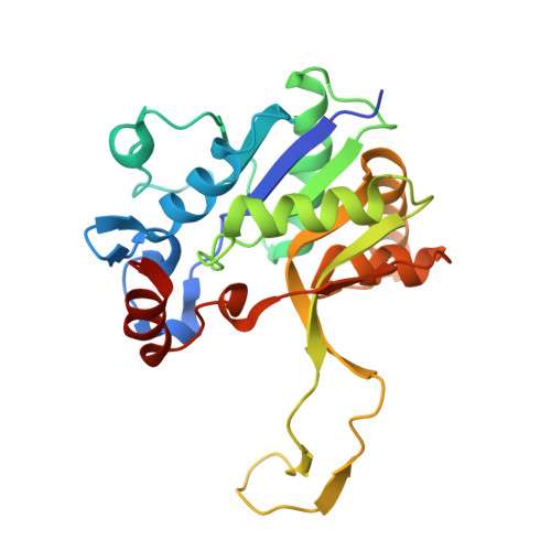
    The methylerythritol phosphate (MEP) pathway is a metabolic pathway that produces the isoprenoids isopentyl pyrophosphate and dimethylallyl pyrophosphate. Notably, the MEP pathway is present in bacteria and not in mammals, which makes the enzymes of the MEP pathway attractive targets for discovering new anti-infective agents due to the reduced chances of off-target interactions leading to side effects. There are seven enzymes in the MEP pathway, the third of which is IspD. Two crystal structures of Burkholderia thailandensis IspD (BtIspD) were determined: an apo structure and that of a complex with cytidine triphosphate (CTP). Comparison of the CTP-bound BtIspD structure with the apo structure revealed that CTP binding stabilizes the loop composed of residues 13-19. The apo structure of Mycobacterium paratuberculosis IspD (MpIspD) is also reported. The melting temperatures of MpIspD and BtIspD were evaluated by circular dichroism. The moderate T m values suggest that a thermal shift assay may be feasible for future inhibitor screening. Finally, the binding affinity of CTP for BtIspD was evaluated by isothermal titration calorimetry. These structural and biophysical data will aid in the discovery of IspD inhibitors.


  • Organizational Affiliation
    • Seattle Structural Genomics Center for Infectious Disease (SSGCID), Seattle, WA 98109, USA.

Macromolecule Content 

  • Total Structure Weight: 105.12 kDa 
  • Atom Count: 6,966 
  • Modeled Residue Count: 898 
  • Deposited Residue Count: 964 
  • Unique protein chains: 1

Macromolecules

Find similar proteins by:|  3D Structure
Entity ID: 1
MoleculeChains  Sequence LengthOrganismDetailsImage
2-C-methyl-D-erythritol 4-phosphate cytidylyltransferase
A, B, C, D
241Burkholderia thailandensis E264Mutation(s): 0 
Gene Names: ispDBTH_I2089
EC: 2.7.7.60
UniProt
Find proteins for Q2SWT6 (Burkholderia thailandensis (strain ATCC 700388 / DSM 13276 / CCUG 48851 / CIP 106301 / E264))
Explore Q2SWT6 
Go to UniProtKB:  Q2SWT6
Entity Groups
Sequence Clusters30% Identity50% Identity70% Identity90% Identity95% Identity100% Identity
UniProt GroupQ2SWT6
Sequence Annotations
Expand
Reference Sequence

Small Molecules

Ligands 5 Unique
IDChains Name / Formula / InChI Key2D Diagram3D Interactions
CTP

Query on CTP


Download:Ideal Coordinates CCD File
E [auth A],
P [auth D]
CYTIDINE-5'-TRIPHOSPHATE
C9 H16 N3 O14 P3
PCDQPRRSZKQHHS-XVFCMESISA-N
CAD

Query on CAD


Download:Ideal Coordinates CCD File
O [auth C]CACODYLIC ACID
C2 H7 As O2
OGGXGZAMXPVRFZ-UHFFFAOYSA-N
GOL

Query on GOL


Download:Ideal Coordinates CCD File
T [auth D]GLYCEROL
C3 H8 O3
PEDCQBHIVMGVHV-UHFFFAOYSA-N
ACT

Query on ACT


Download:Ideal Coordinates CCD File
G [auth A]
H [auth A]
I [auth A]
J [auth A]
K [auth A]
G [auth A],
H [auth A],
I [auth A],
J [auth A],
K [auth A],
L [auth C],
M [auth C],
N [auth C],
R [auth D],
S [auth D]
ACETATE ION
C2 H3 O2
QTBSBXVTEAMEQO-UHFFFAOYSA-M
MG

Query on MG


Download:Ideal Coordinates CCD File
F [auth A],
Q [auth D]
MAGNESIUM ION
Mg
JLVVSXFLKOJNIY-UHFFFAOYSA-N

Experimental Data & Validation

Experimental Data

  • Method: X-RAY DIFFRACTION
  • Resolution: 2.30 Å
  • R-Value Free:  0.257 (Depositor), 0.258 (DCC) 
  • R-Value Work:  0.205 (Depositor), 0.206 (DCC) 
  • R-Value Observed: 0.207 (Depositor) 
Space Group: P 1 21 1
Unit Cell:
Length ( Å )Angle ( ˚ )
a = 45.9α = 90
b = 138.95β = 101.94
c = 76.92γ = 90
Software Package:
Software NamePurpose
XSCALEdata scaling
PHENIXrefinement
PDB_EXTRACTdata extraction
XDSdata reduction
PHENIXphasing

Structure Validation

View Full Validation Report


Ligand Structure Quality Assessment 


Entry History 

Deposition Data

Revision History  (Full details and data files)

  • Version 1.0: 2015-04-29
    Type: Initial release
  • Version 1.1: 2015-05-06
    Changes: Derived calculations
  • Version 1.2: 2017-10-11
    Changes: Data collection, Derived calculations, Source and taxonomy
  • Version 1.3: 2023-09-27
    Changes: Data collection, Database references, Refinement description
  • Version 1.4: 2024-02-14
    Changes: Database references