4ZCW | pdb_00004zcw

Structure of Human Enolase 2 in complex with SF2312


Experimental Data Snapshot

  • Method: X-RAY DIFFRACTION
  • Resolution: 1.99 Å
  • R-Value Free: 
    0.202 (Depositor), 0.204 (DCC) 
  • R-Value Work: 
    0.157 (Depositor), 0.159 (DCC) 
  • R-Value Observed: 
    0.159 (Depositor) 

Starting Model: experimental
View more details

wwPDB Validation 3D Report Full Report

Validation slider image for 4ZCW

Ligand Structure Quality Assessment 


This is version 1.5 of the entry. See complete history

Literature

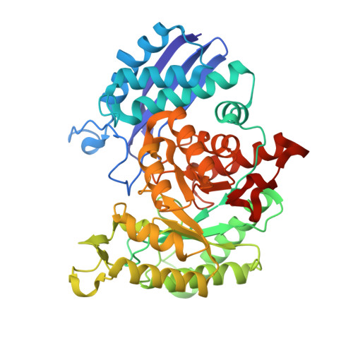
SF2312 is a natural phosphonate inhibitor of enolase.

Leonard, P.G.Satani, N.Maxwell, D.Lin, Y.H.Hammoudi, N.Peng, Z.Pisaneschi, F.Link, T.M.Lee, G.R.Sun, D.Prasad, B.A.Di Francesco, M.E.Czako, B.Asara, J.M.Wang, Y.A.Bornmann, W.DePinho, R.A.Muller, F.L.

(2016) Nat Chem Biol 12: 1053-1058

  • DOI: https://doi.org/10.1038/nchembio.2195
  • Primary Citation Related Structures: 
    4ZA0, 4ZCW

  • PubMed Abstract: 

    Despite being crucial for energy generation in most forms of life, few if any microbial antibiotics specifically inhibit glycolysis. To develop a specific inhibitor of the glycolytic enzyme enolase 2 (ENO2) for the treatment of cancers with deletion of ENO1 (encoding enolase 1), we modeled the synthetic tool compound inhibitor phosphonoacetohydroxamate (PhAH) into the active site of human ENO2. A ring-stabilized analog of PhAH, in which the hydroxamic nitrogen is linked to Cα by an ethylene bridge, was predicted to increase binding affinity by stabilizing the inhibitor in a bound conformation. Unexpectedly, a structure-based search revealed that our hypothesized backbone-stabilized PhAH bears strong similarity to SF2312, a phosphonate antibiotic of unknown mode of action produced by the actinomycete Micromonospora, which is active under anaerobic conditions. Here, we present multiple lines of evidence, including a novel X-ray structure, that SF2312 is a highly potent, low-nanomolar inhibitor of enolase.


  • Organizational Affiliation
    • Department of Genomic Medicine and Core for Biomolecular Structure and Function, University of Texas MD Anderson Cancer Center, Houston, TX 77054.

Macromolecule Content 

  • Total Structure Weight: 96.79 kDa 
  • Atom Count: 7,276 
  • Modeled Residue Count: 866 
  • Deposited Residue Count: 880 
  • Unique protein chains: 1

Macromolecules

Find similar proteins by:|  3D Structure
Entity ID: 1
MoleculeChains  Sequence LengthOrganismDetailsImage
Gamma-enolase
A, B
440Homo sapiensMutation(s): 0 
Gene Names: ENO2
EC: 4.2.1.11
UniProt & NIH Common Fund Data Resources
Find proteins for P09104 (Homo sapiens)
Explore P09104 
Go to UniProtKB:  P09104
PHAROS:  P09104
GTEx:  ENSG00000111674 
Entity Groups
Sequence Clusters30% Identity50% Identity70% Identity90% Identity95% Identity100% Identity
UniProt GroupP09104
Sequence Annotations
Expand
Reference Sequence

Experimental Data & Validation

Experimental Data

  • Method: X-RAY DIFFRACTION
  • Resolution: 1.99 Å
  • R-Value Free:  0.202 (Depositor), 0.204 (DCC) 
  • R-Value Work:  0.157 (Depositor), 0.159 (DCC) 
  • R-Value Observed: 0.159 (Depositor) 
Space Group: P 21 21 21
Unit Cell:
Length ( Å )Angle ( ˚ )
a = 68.3α = 90
b = 109.46β = 90
c = 117.27γ = 90
Software Package:
Software NamePurpose
Aimlessdata scaling
PHASERphasing
PHENIXrefinement
Cootmodel building
PDB_EXTRACTdata extraction
iMOSFLMdata reduction

Structure Validation

View Full Validation Report


Ligand Structure Quality Assessment 


Entry History 

Deposition Data

Revision History  (Full details and data files)

  • Version 1.0: 2016-07-20
    Type: Initial release
  • Version 1.1: 2016-10-05
    Changes: Database references
  • Version 1.2: 2016-10-26
    Changes: Database references
  • Version 1.3: 2016-11-23
    Changes: Database references
  • Version 1.4: 2017-11-22
    Changes: Refinement description
  • Version 1.5: 2023-09-27
    Changes: Data collection, Database references, Derived calculations, Refinement description