4Z86 | pdb_00004z86

Crystal structure of Peptidyl-tRNA hydrolase mutant -N118D from Vibrio cholerae at 1.63A resolution.


Experimental Data Snapshot

  • Method: X-RAY DIFFRACTION
  • Resolution: 1.63 Å
  • R-Value Free: 
    0.207 (Depositor), 0.188 (DCC) 
  • R-Value Work: 
    0.170 (Depositor), 0.185 (DCC) 
  • R-Value Observed: 
    0.172 (Depositor) 

wwPDB Validation 3D Report Full Report

Validation slider image for 4Z86

This is version 1.2 of the entry. See complete history

Literature

Unraveling the stereochemical and dynamic aspects of the catalytic site of bacterial peptidyl-tRNA hydrolase.

Kabra, A.Shahid, S.Pal, R.K.Yadav, R.Pulavarti, S.V.Jain, A.Tripathi, S.Arora, A.

(2017) RNA 23: 202-216

  • DOI: https://doi.org/10.1261/rna.057620.116
  • Primary Citation Related Structures: 
    4Z86, 4ZXP, 5B6J, 5IKE, 5IMB

  • PubMed Abstract: 

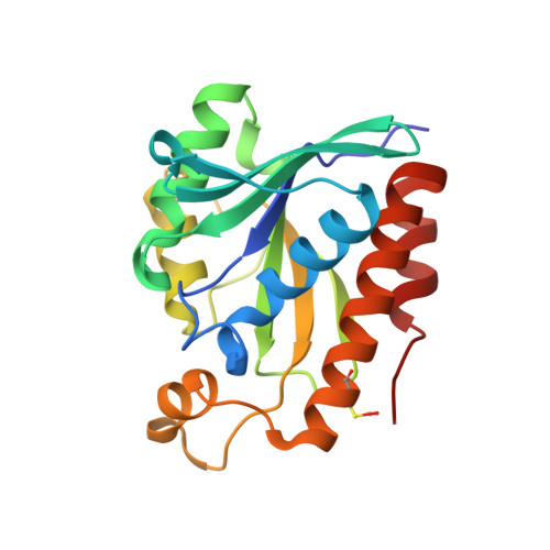
    Bacterial peptidyl-tRNA hydrolase (Pth; EC 3.1.1.29) hydrolyzes the peptidyl-tRNAs accumulated in the cytoplasm and thereby prevents cell death by alleviating tRNA starvation. X-ray and NMR studies of Vibrio cholerae Pth (VcPth) and mutants of its key residues involved in catalysis show that the activity and selectivity of the protein depends on the stereochemistry and dynamics of residues H24, D97, N118, and N14. D97-H24 interaction is critical for activity because it increases the nucleophilicity of H24. The N118 and N14 have orthogonally competing interactions with H24, both of which reduce the nucleophilicity of H24 and are likely to be offset by positioning of a peptidyl-tRNA substrate. The region proximal to H24 and the lid region exhibit slow motions that may assist in accommodating the substrate. Helix α3 exhibits a slow wobble with intermediate time scale motions of its N-cap residue N118, which may work as a flypaper to position the scissile ester bond of the substrate. Overall, the dynamics of interactions between the side chains of N14, H24, D97, and N118, control the catalysis of substrate by this enzyme.


  • Organizational Affiliation
    • Molecular and Structural Biology Division, CSIR-Central Drug Research Institute, Lucknow 226031, India.

Macromolecule Content 

  • Total Structure Weight: 43.55 kDa 
  • Atom Count: 3,297 
  • Modeled Residue Count: 392 
  • Deposited Residue Count: 398 
  • Unique protein chains: 1

Macromolecules

Find similar proteins by:|  3D Structure
Entity ID: 1
MoleculeChains  Sequence LengthOrganismDetailsImage
Peptidyl-tRNA hydrolase
A, B
199Vibrio cholerae O1 biovar El Tor str. N16961Mutation(s): 1 
Gene Names: pthVC_2184
EC: 3.1.1.29
UniProt
Find proteins for Q9KQ21 (Vibrio cholerae serotype O1 (strain ATCC 39315 / El Tor Inaba N16961))
Explore Q9KQ21 
Go to UniProtKB:  Q9KQ21
Entity Groups
Sequence Clusters30% Identity50% Identity70% Identity90% Identity95% Identity100% Identity
UniProt GroupQ9KQ21
Sequence Annotations
Expand
Reference Sequence

Small Molecules

Modified Residues  1 Unique
IDChains TypeFormula2D DiagramParent
2CO
Query on 2CO
A, B
L-PEPTIDE LINKINGC3 H7 N O4 SCYS

Experimental Data & Validation

Experimental Data

  • Method: X-RAY DIFFRACTION
  • Resolution: 1.63 Å
  • R-Value Free:  0.207 (Depositor), 0.188 (DCC) 
  • R-Value Work:  0.170 (Depositor), 0.185 (DCC) 
  • R-Value Observed: 0.172 (Depositor) 
Space Group: P 21 21 21
Unit Cell:
Length ( Å )Angle ( ˚ )
a = 44.112α = 90
b = 71.124β = 90
c = 124.03γ = 90
Software Package:
Software NamePurpose
REFMACrefinement
HKL-2000data reduction
HKL-2000data scaling
HKL-2000phasing

Structure Validation

View Full Validation Report


Entry History 

Deposition Data

Revision History  (Full details and data files)

  • Version 1.0: 2016-06-08
    Type: Initial release
  • Version 1.1: 2017-04-05
    Changes: Database references
  • Version 1.2: 2024-11-13
    Changes: Data collection, Database references, Structure summary