4YU5 | pdb_00004yu5

Crystal structure of selenomethionine variant of Bacillus anthracis immune inhibitor A2 peptidase zymogen


Experimental Data Snapshot

  • Method: X-RAY DIFFRACTION
  • Resolution: 2.90 Å
  • R-Value Free: 
    0.207 (Depositor), 0.216 (DCC) 
  • R-Value Work: 
    0.182 (Depositor), 0.188 (DCC) 
  • R-Value Observed: 
    0.183 (Depositor) 

wwPDB Validation 3D Report Full Report

Validation slider image for 4YU5

Ligand Structure Quality Assessment 


This is version 1.2 of the entry. See complete history

Literature

Structural Basis for Latency and Function of Immune Inhibitor A Metallopeptidase, a Modulator of the Bacillus anthracis Secretome.

Arolas, J.L.Goulas, T.Pomerantsev, A.P.Leppla, S.H.Gomis-Ruth, F.X.

(2016) Structure 24: 25-36

  • DOI: https://doi.org/10.1016/j.str.2015.10.015
  • Primary Citation Related Structures: 
    4YU5, 4YU6

  • PubMed Abstract: 

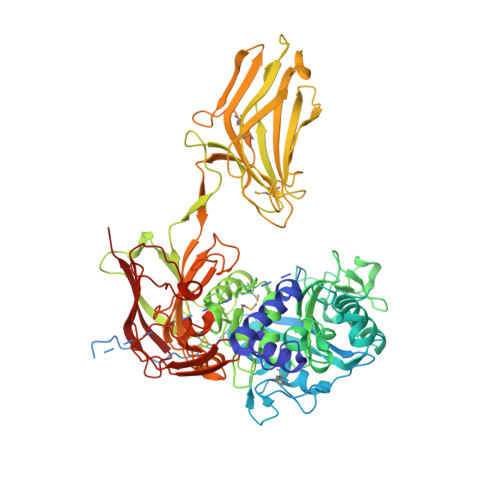
    Immune inhibitor A(InhA)-type metallopeptidases are potential virulence factors secreted by members of the Bacillus cereus group. Two paralogs from anthrax-causing Bacillus anthracis (BaInhA1 and BaInhA2) were shown to degrade host tissue proteins with broad substrate specificity. Analysis of their activation mechanism and the crystal structure of a zymogenic BaInhA2 variant revealed a ∼750-residue four-domain structure featuring a pro-peptide, a catalytic domain, a domain reminiscent of viral envelope glycoproteins, and a MAM domain grafted into the latter. This domain, previously found only in eukaryotes, is required for proper protein expression in B. anthracis and evinces certain flexibility. Latency is uniquely modulated by the N-terminal segment of the pro-peptide, which binds the catalytic zinc through its α-amino group and occupies the primed side of the active-site cleft. The present results further our understanding of the modus operandi of an anthrax secretome regulator.


  • Organizational Affiliation
    • Proteolysis Lab, Department of Structural Biology ("María de Maeztu" Unit of Excellence), Molecular Biology Institute of Barcelona, Spanish Research Council (CSIC), Barcelona Science Park, Helix Building, Baldiri Reixac, 15-21, 08028 Barcelona, Spain.

Macromolecule Content 

  • Total Structure Weight: 169.73 kDa 
  • Atom Count: 10,942 
  • Modeled Residue Count: 1,384 
  • Deposited Residue Count: 1,512 
  • Unique protein chains: 1

Macromolecules

Find similar proteins by:|  3D Structure
Entity ID: 1
MoleculeChains  Sequence LengthOrganismDetailsImage
Immune inhibitor A, metalloprotease
A, B
756Bacillus cereus biovar anthracis str. CIMutation(s): 1 
Gene Names: inhA2BACI_c06810
EC: 3.4.24

Small Molecules

Ligands 5 Unique
IDChains Name / Formula / InChI Key2D Diagram3D Interactions
KH2

Query on KH2


Download:Ideal Coordinates CCD File
H [auth A],
I [auth A]
3-(1-methylpiperidinium-1-yl)propane-1-sulfonate
C9 H19 N O3 S
DQNQWAVIDNVATL-UHFFFAOYSA-N
GOL

Query on GOL


Download:Ideal Coordinates CCD File
J [auth A]
K [auth A]
L [auth A]
M [auth A]
N [auth A]
J [auth A],
K [auth A],
L [auth A],
M [auth A],
N [auth A],
O [auth A]
GLYCEROL
C3 H8 O3
PEDCQBHIVMGVHV-UHFFFAOYSA-N
ZN

Query on ZN


Download:Ideal Coordinates CCD File
C [auth A],
P [auth B]
ZINC ION
Zn
PTFCDOFLOPIGGS-UHFFFAOYSA-N
CA

Query on CA


Download:Ideal Coordinates CCD File
D [auth A]
E [auth A]
F [auth A]
G [auth A]
Q [auth B]
D [auth A],
E [auth A],
F [auth A],
G [auth A],
Q [auth B],
R [auth B],
S [auth B],
T [auth B]
CALCIUM ION
Ca
BHPQYMZQTOCNFJ-UHFFFAOYSA-N
K

Query on K


Download:Ideal Coordinates CCD File
U [auth B]POTASSIUM ION
K
NPYPAHLBTDXSSS-UHFFFAOYSA-N
Modified Residues  1 Unique
IDChains TypeFormula2D DiagramParent
MSE
Query on MSE
A, B
L-PEPTIDE LINKINGC5 H11 N O2 SeMET

Experimental Data & Validation

Experimental Data

  • Method: X-RAY DIFFRACTION
  • Resolution: 2.90 Å
  • R-Value Free:  0.207 (Depositor), 0.216 (DCC) 
  • R-Value Work:  0.182 (Depositor), 0.188 (DCC) 
  • R-Value Observed: 0.183 (Depositor) 
Space Group: P 21 21 21
Unit Cell:
Length ( Å )Angle ( ˚ )
a = 97.61α = 90
b = 102.41β = 90
c = 242.88γ = 90
Software Package:
Software NamePurpose
XDSdata reduction
XSCALEdata scaling
Cootmodel building
SHELXphasing
BUSTERrefinement

Structure Validation

View Full Validation Report


Ligand Structure Quality Assessment 


Entry History 

Deposition Data

Revision History  (Full details and data files)

  • Version 1.0: 2015-10-28
    Type: Initial release
  • Version 1.1: 2016-01-20
    Changes: Database references
  • Version 1.2: 2024-11-06
    Changes: Data collection, Database references, Derived calculations, Structure summary