4YU0 | pdb_00004yu0

Crystal structure of a tetramer of GluA2 TR mutant ligand binding domains bound with glutamate at 1.26 Angstrom resolution


Experimental Data Snapshot

  • Method: X-RAY DIFFRACTION
  • Resolution: 1.26 Å
  • R-Value Free: 
    0.157 (Depositor), 0.158 (DCC) 
  • R-Value Work: 
    0.129 (Depositor), 0.131 (DCC) 
  • R-Value Observed: 
    0.131 (Depositor) 

Starting Model: experimental
View more details

wwPDB Validation 3D Report Full Report

Validation slider image for 4YU0

This is version 2.2 of the entry. See complete history

Literature

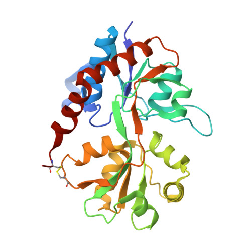
Crystal structure of the tetrameric GluA2 ligand-binding domain in complex with glutamate at 1.26 Angstroms resolution

Chebli, M.Salazar, H.Baranovic, J.Carbone, A.L.Ghisi, V.Faelber, K.Lau, A.Y.Daumke, O.Plested, A.J.R.

To be published.

Macromolecule Content 

  • Total Structure Weight: 59.66 kDa 
  • Atom Count: 5,112 
  • Modeled Residue Count: 518 
  • Deposited Residue Count: 526 
  • Unique protein chains: 1

Macromolecules

Find similar proteins by:|  3D Structure
Entity ID: 1
MoleculeChains  Sequence LengthOrganismDetailsImage
Glutamate receptor 2,Glutamate receptor 2
A, B
263Rattus norvegicusMutation(s): 2 
Gene Names: Gria2Glur2
UniProt
Find proteins for P19491 (Rattus norvegicus)
Explore P19491 
Go to UniProtKB:  P19491
Entity Groups
Sequence Clusters30% Identity50% Identity70% Identity90% Identity95% Identity100% Identity
UniProt GroupP19491
Sequence Annotations
Expand
Reference Sequence

Small Molecules

Ligands 3 Unique
IDChains Name / Formula / InChI Key2D Diagram3D Interactions
GLU

Query on GLU


Download:Ideal Coordinates CCD File
C [auth A],
I [auth B]
GLUTAMIC ACID
C5 H9 N O4
WHUUTDBJXJRKMK-VKHMYHEASA-N
PEG

Query on PEG


Download:Ideal Coordinates CCD File
G [auth A],
H [auth A],
M [auth B],
N [auth B]
DI(HYDROXYETHYL)ETHER
C4 H10 O3
MTHSVFCYNBDYFN-UHFFFAOYSA-N
PO4

Query on PO4


Download:Ideal Coordinates CCD File
D [auth A]
E [auth A]
F [auth A]
J [auth B]
K [auth B]
D [auth A],
E [auth A],
F [auth A],
J [auth B],
K [auth B],
L [auth B]
PHOSPHATE ION
O4 P
NBIIXXVUZAFLBC-UHFFFAOYSA-K

Experimental Data & Validation

Experimental Data

  • Method: X-RAY DIFFRACTION
  • Resolution: 1.26 Å
  • R-Value Free:  0.157 (Depositor), 0.158 (DCC) 
  • R-Value Work:  0.129 (Depositor), 0.131 (DCC) 
  • R-Value Observed: 0.131 (Depositor) 
Space Group: P 1 2 1
Unit Cell:
Length ( Å )Angle ( ˚ )
a = 47.14α = 90
b = 47.42β = 93.59
c = 116.94γ = 90
Software Package:
Software NamePurpose
PHENIXrefinement
MOSFLMdata reduction
PHASERphasing
Cootmodel building
XDSdata scaling

Structure Validation

View Full Validation Report


Entry History 

Deposition Data

Revision History  (Full details and data files)

  • Version 1.0: 2016-01-13
    Type: Initial release
  • Version 2.0: 2021-03-31
    Changes: Advisory, Atomic model, Data collection, Derived calculations
  • Version 2.1: 2024-01-10
    Changes: Data collection, Database references, Refinement description
  • Version 2.2: 2024-11-13
    Changes: Structure summary