4YRV

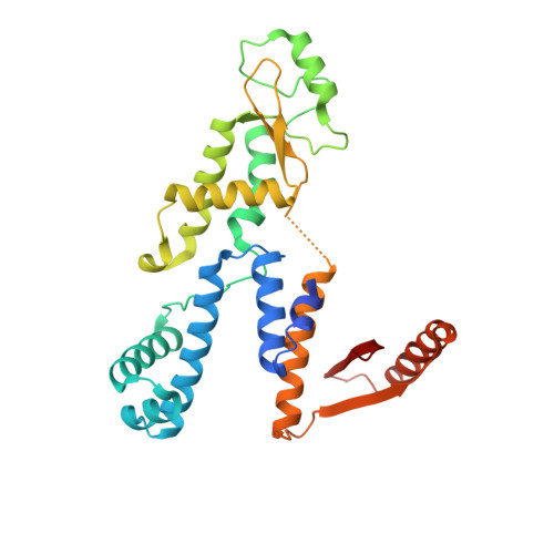
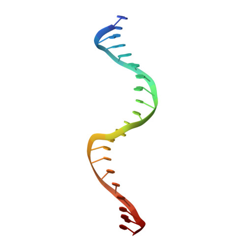
Crystal structure of Anabaena transcription factor HetR complexed with 21-bp DNA from hetP promoter


Experimental Data Snapshot

  • Method: X-RAY DIFFRACTION
  • Resolution: 2.80 Å
  • R-Value Free: 0.264 
  • R-Value Work: 0.203 
  • R-Value Observed: 0.206 

Starting Model: experimental
View more details

wwPDB Validation   3D Report Full Report


This is version 1.1 of the entry. See complete history


Literature

Structural insights into HetR-PatS interaction involved in cyanobacterial pattern formation

Hu, H.X.Jiang, Y.L.Zhao, M.X.Cai, K.Liu, S.Wen, B.Lv, P.Zhang, Y.Peng, J.Zhong, H.Yu, H.M.Ren, Y.M.Zhang, Z.Tian, C.Wu, Q.Oliveberg, M.Zhang, C.C.Chen, Y.Zhou, C.Z.

(2015) Sci Rep 5: 16470-16470

  • DOI: https://doi.org/10.1038/srep16470
  • Primary Citation of Related Structures:  
    4YNL, 4YRV

  • PubMed Abstract: 

    The one-dimensional pattern of heterocyst in the model cyanobacterium Anabaena sp. PCC 7120 is coordinated by the transcription factor HetR and PatS peptide. Here we report the complex structures of HetR binding to DNA, and its hood domain (HetRHood) binding to a PatS-derived hexapeptide (PatS6) at 2.80 and 2.10 Å, respectively. The intertwined HetR dimer possesses a couple of novel HTH motifs, each of which consists of two canonical α-helices in the DNA-binding domain and an auxiliary α-helix from the flap domain of the neighboring subunit. Two PatS6 peptides bind to the lateral clefts of HetRHood, and trigger significant conformational changes of the flap domain, resulting in dissociation of the auxiliary α-helix and eventually release of HetR from the DNA major grove. These findings provide the structural insights into a prokaryotic example of Turing model.


  • Organizational Affiliation

    Hefei National Laboratory for Physical Sciences at the Microscale and School of Life Sciences, University of Science and Technology of China, Hefei Anhui 230027, China.


Macromolecules

Find similar proteins by:  (by identity cutoff)  |  3D Structure
Entity ID: 1
MoleculeChains Sequence LengthOrganismDetailsImage
Heterocyst differentiation control proteinA [auth B],
B [auth A]
307Nostoc sp. PCC 7120 = FACHB-418Mutation(s): 0 
Gene Names: hetRalr2339
EC: 3.4.21
UniProt
Find proteins for P27709 (Nostoc sp. (strain PCC 7120 / SAG 25.82 / UTEX 2576))
Explore P27709 
Go to UniProtKB:  P27709
Entity Groups  
Sequence Clusters30% Identity50% Identity70% Identity90% Identity95% Identity100% Identity
UniProt GroupP27709
Sequence Annotations
Expand
  • Reference Sequence

Find similar nucleic acids by:  Sequence   |   3D Structure  

Entity ID: 2
MoleculeChains LengthOrganismImage
DNA (5'-D(P*GP*CP*GP*AP*GP*GP*GP*GP*TP*CP*TP*AP*AP*CP*CP*CP*CP*TP*CP*AP*T)-3')21Nostoc sp. PCC 7120 = FACHB-418
Sequence Annotations
Expand
  • Reference Sequence

Find similar nucleic acids by:  Sequence   |   3D Structure  

Entity ID: 3
MoleculeChains LengthOrganismImage
DNA (5'-D(P*AP*TP*GP*AP*GP*GP*GP*GP*TP*TP*AP*GP*AP*CP*CP*CP*CP*TP*CP*GP*C)-3')21Nostoc sp. PCC 7120 = FACHB-418
Sequence Annotations
Expand
  • Reference Sequence
Experimental Data & Validation

Experimental Data

  • Method: X-RAY DIFFRACTION
  • Resolution: 2.80 Å
  • R-Value Free: 0.264 
  • R-Value Work: 0.203 
  • R-Value Observed: 0.206 
  • Space Group: P 41 21 2
Unit Cell:
Length ( Å )Angle ( ˚ )
a = 90.559α = 90
b = 90.559β = 90
c = 242.32γ = 90
Software Package:
Software NamePurpose
PHENIXrefinement
HKL-2000data reduction
HKL-2000data scaling
MOLREPphasing

Structure Validation

View Full Validation Report


Entry History & Funding Information

Deposition Data


Funding OrganizationLocationGrant Number
National Natural Science Foundation of ChinaChina31370757
National Natural Science Foundation of ChinaChina31070652

Revision History  (Full details and data files)

  • Version 1.0: 2015-12-02
    Type: Initial release
  • Version 1.1: 2023-11-08
    Changes: Data collection, Database references, Derived calculations, Refinement description