4YIR | pdb_00004yir

Crystal structure of Rad4-Rad23 crosslinked to an undamaged DNA


Experimental Data Snapshot

  • Method: X-RAY DIFFRACTION
  • Resolution: 3.05 Å
  • R-Value Free: 
    0.253 (Depositor), 0.254 (DCC) 
  • R-Value Work: 
    0.192 (Depositor), 0.198 (DCC) 
  • R-Value Observed: 
    0.197 (Depositor) 

Starting Model: experimental
View more details

wwPDB Validation 3D Report Full Report

Validation slider image for 4YIR

This is version 1.2 of the entry. See complete history

Literature

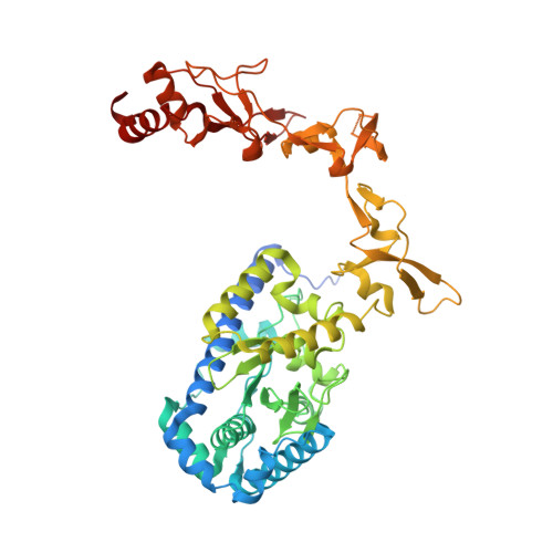
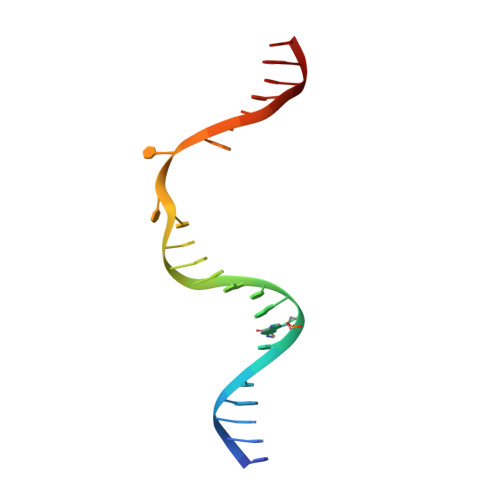
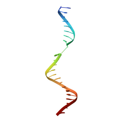
Kinetic gating mechanism of DNA damage recognition by Rad4/XPC.

Chen, X.Velmurugu, Y.Zheng, G.Park, B.Shim, Y.Kim, Y.Liu, L.Van Houten, B.He, C.Ansari, A.Min, J.H.

(2015) Nat Commun 6: 5849

  • DOI: https://doi.org/10.1038/ncomms6849
  • Primary Citation Related Structures: 
    4YIR, 6UBF

  • PubMed Abstract: 

    The xeroderma pigmentosum C (XPC) complex initiates nucleotide excision repair by recognizing DNA lesions before recruiting downstream factors. How XPC detects structurally diverse lesions embedded within normal DNA is unknown. Here we present a crystal structure that captures the yeast XPC orthologue (Rad4) on a single register of undamaged DNA. The structure shows that a disulphide-tethered Rad4 flips out normal nucleotides and adopts a conformation similar to that seen with damaged DNA. Contrary to many DNA repair enzymes that can directly reject non-target sites as structural misfits, our results suggest that Rad4/XPC uses a kinetic gating mechanism whereby lesion selectivity arises from the kinetic competition between DNA opening and the residence time of Rad4/XPC per site. This mechanism is further supported by measurements of Rad4-induced lesion-opening times using temperature-jump perturbation spectroscopy. Kinetic gating may be a general mechanism used by site-specific DNA-binding proteins to minimize time-consuming interrogations of non-target sites.


  • Organizational Affiliation
    • Department of Chemistry, University of Illinois at Chicago, 845 W. Taylor Street, Chicago, Illinois 60607, USA.

Macromolecule Content 

  • Total Structure Weight: 95.17 kDa 
  • Atom Count: 5,459 
  • Modeled Residue Count: 599 
  • Deposited Residue Count: 757 
  • Unique protein chains: 2
  • Unique nucleic acid chains: 2

Macromolecules


Find similar proteins by:|  3D Structure
Entity ID: 1
MoleculeChains  Sequence LengthOrganismDetailsImage
DNA repair protein RAD4538Saccharomyces cerevisiae S288CMutation(s): 2 
Gene Names: RAD4YER162C
UniProt
Find proteins for P14736 (Saccharomyces cerevisiae (strain ATCC 204508 / S288c))
Explore P14736 
Go to UniProtKB:  P14736
Entity Groups
Sequence Clusters30% Identity50% Identity70% Identity90% Identity95% Identity100% Identity
UniProt GroupP14736
Sequence Annotations
Expand
Reference Sequence
Find similar proteins by:|  3D Structure
Entity ID: 2
MoleculeChains  Sequence LengthOrganismDetailsImage
UV excision repair protein RAD23B [auth X]171Saccharomyces cerevisiae S288CMutation(s): 0 
Gene Names: RAD23YEL037CSYGP-ORF29
UniProt
Find proteins for P32628 (Saccharomyces cerevisiae (strain ATCC 204508 / S288c))
Explore P32628 
Go to UniProtKB:  P32628
Entity Groups
Sequence Clusters30% Identity50% Identity70% Identity90% Identity95% Identity100% Identity
UniProt GroupP32628
Sequence Annotations
Expand
Reference Sequence
Find similar nucleic acids by:  Sequence
Entity ID: 3
MoleculeChains LengthOrganismImage
DNA (5'-D(*TP*TP*GP*AP*CP*TP*CP*(G47)P*AP*CP*AP*TP*CP*CP*CP*CP*CP*GP*CP*TP*AP*CP*AP*A)-3')C [auth W]24synthetic construct
Sequence Annotations
Expand
Reference Sequence
Find similar nucleic acids by:  Sequence
Entity ID: 4
MoleculeChains LengthOrganismImage
DNA (5'-D(*AP*TP*TP*GP*TP*AP*GP*CP*G*GP*GP*GP*AP*TP*GP*TP*CP*GP*AP*GP*TP*CP*A)-3')D [auth Y]24synthetic construct
Sequence Annotations
Expand
Reference Sequence

Experimental Data & Validation

Experimental Data

  • Method: X-RAY DIFFRACTION
  • Resolution: 3.05 Å
  • R-Value Free:  0.253 (Depositor), 0.254 (DCC) 
  • R-Value Work:  0.192 (Depositor), 0.198 (DCC) 
  • R-Value Observed: 0.197 (Depositor) 
Space Group: P 41 21 2
Unit Cell:
Length ( Å )Angle ( ˚ )
a = 79.405α = 90
b = 79.405β = 90
c = 404.366γ = 90
Software Package:
Software NamePurpose
PHENIXrefinement
HKL-3000data reduction
HKL-3000data scaling
PHENIXphasing

Structure Validation

View Full Validation Report


Entry History 

& Funding Information

Deposition Data


Funding OrganizationLocationGrant Number
The Searle Funds at The Chicago Community TrustUnited States--
The Chancellor's Discovery Fund, University of Illinois at ChicagoUnited States--
Startup funding, University of Illinois at ChicagoUnited States--

Revision History  (Full details and data files)

  • Version 1.0: 2015-03-11
    Type: Initial release
  • Version 1.1: 2023-09-27
    Changes: Data collection, Database references, Derived calculations, Refinement description, Source and taxonomy
  • Version 1.2: 2024-10-23
    Changes: Structure summary