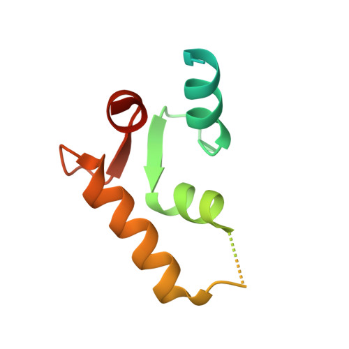
4YGB

Crystal structure of ERGIC-53/MCFD2, monoclinic calcium-free form


Experimental Data Snapshot

  • Method: X-RAY DIFFRACTION
  • Resolution: 1.60 Å
  • R-Value Free: 0.199 
  • R-Value Work: 0.180 
  • R-Value Observed: 0.181 

Starting Model: experimental
View more details

wwPDB Validation   3D Report Full Report


This is version 1.4 of the entry. See complete history


Literature

Crystallographic snapshots of the EF-hand protein MCFD2 complexed with the intracellular lectin ERGIC-53 involved in glycoprotein transport.

Satoh, T.Nishio, M.Suzuki, K.Yagi-Utsumi, M.Kamiya, Y.Mizushima, T.Kato, K.

(2020) Acta Crystallogr F Struct Biol Commun 76: 216-221

  • DOI: https://doi.org/10.1107/S2053230X20005452
  • Primary Citation of Related Structures:  
    4YGB, 4YGC, 4YGD, 4YGE

  • PubMed Abstract: 

    The transmembrane intracellular lectin ER-Golgi intermediate compartment protein 53 (ERGIC-53) and the soluble EF-hand multiple coagulation factor deficiency protein 2 (MCFD2) form a complex that functions as a cargo receptor, trafficking various glycoproteins between the endoplasmic reticulum (ER) and the Golgi apparatus. It has been demonstrated that the carbohydrate-recognition domain (CRD) of ERGIC-53 (ERGIC-53 CRD ) interacts with N-linked glycans on cargo glycoproteins, whereas MCFD2 recognizes polypeptide segments of cargo glycoproteins. Crystal structures of ERGIC-53 CRD complexed with MCFD2 and mannosyl oligosaccharides have revealed protein-protein and protein-sugar binding modes. In contrast, the polypeptide-recognition mechanism of MCFD2 remains largely unknown. Here, a 1.60 Å resolution crystal structure of the ERGIC-53 CRD -MCFD2 complex is reported, along with three other crystal forms. Comparison of these structures with those previously reported reveal that MCFD2, but not ERGIC-53-CRD, exhibits significant conformational plasticity that may be relevant to its accommodation of various polypeptide ligands.


  • Organizational Affiliation

    Graduate School of Pharmaceutical Sciences, Nagoya City University, 3-1 Tanabe-dori, Mizuho-ku, Nagoya, Aichi 467-8603, Japan.


Macromolecules
Find similar proteins by:  (by identity cutoff)  |  3D Structure
Entity ID: 1
MoleculeChains Sequence LengthOrganismDetailsImage
Protein ERGIC-53
A, C
246Homo sapiensMutation(s): 0 
Gene Names: LMAN1ERGIC53F5F8D
UniProt & NIH Common Fund Data Resources
Find proteins for P49257 (Homo sapiens)
Explore P49257 
Go to UniProtKB:  P49257
PHAROS:  P49257
GTEx:  ENSG00000074695 
Entity Groups  
Sequence Clusters30% Identity50% Identity70% Identity90% Identity95% Identity100% Identity
UniProt GroupP49257
Sequence Annotations
Expand
  • Reference Sequence
Find similar proteins by:  (by identity cutoff)  |  3D Structure
Entity ID: 2
MoleculeChains Sequence LengthOrganismDetailsImage
Multiple coagulation factor deficiency protein 2
B, D
104Homo sapiensMutation(s): 0 
Gene Names: MCFD2SDNSF
UniProt & NIH Common Fund Data Resources
Find proteins for Q8NI22 (Homo sapiens)
Explore Q8NI22 
Go to UniProtKB:  Q8NI22
PHAROS:  Q8NI22
GTEx:  ENSG00000180398 
Entity Groups  
Sequence Clusters30% Identity50% Identity70% Identity90% Identity95% Identity100% Identity
UniProt GroupQ8NI22
Sequence Annotations
Expand
  • Reference Sequence
Experimental Data & Validation

Experimental Data

  • Method: X-RAY DIFFRACTION
  • Resolution: 1.60 Å
  • R-Value Free: 0.199 
  • R-Value Work: 0.180 
  • R-Value Observed: 0.181 
  • Space Group: P 1 21 1
Unit Cell:
Length ( Å )Angle ( ˚ )
a = 57.924α = 90
b = 116.93β = 120.13
c = 58.065γ = 90
Software Package:
Software NamePurpose
REFMACrefinement
DENZOdata reduction
SCALEPACKdata scaling
MOLREPphasing

Structure Validation

View Full Validation Report


Entry History & Funding Information

Deposition Data


Funding OrganizationLocationGrant Number
JSPS KAKENHIJapan25121730, 25102008, 24249002
Okazaki ORION project, Ministry of Education, Culture, Sports, Science and TechnologyJapan--
Nanotechnology Platform Project, Ministry of Education, Culture, Sports, Science and TechnologyJapan--

Revision History  (Full details and data files)

  • Version 1.0: 2016-04-06
    Type: Initial release
  • Version 1.1: 2020-02-05
    Changes: Data collection, Derived calculations
  • Version 1.2: 2020-05-13
    Changes: Database references
  • Version 1.3: 2023-11-08
    Changes: Data collection, Database references, Derived calculations, Refinement description
  • Version 1.4: 2024-10-23
    Changes: Structure summary