4YC2 | pdb_00004yc2

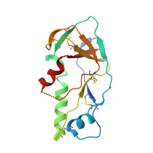
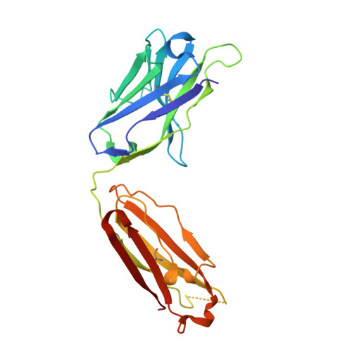
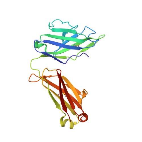
Crystal structure of the stabilized inner domain of clade A/E HIV-1 gp120 from E. coli in complex with the antibody A32.


Experimental Data Snapshot

  • Method: X-RAY DIFFRACTION
  • Resolution: 3.02 Å
  • R-Value Free: 
    0.288 (Depositor), 0.284 (DCC) 
  • R-Value Work: 
    0.227 (Depositor), 0.229 (DCC) 
  • R-Value Observed: 
    0.230 (Depositor) 

Starting Models: experimental
View more details

wwPDB Validation 3D Report Full Report

Validation slider image for 4YC2

This is version 1.6 of the entry. See complete history. 

Literature

Paring Down HIV Env: Design and Crystal Structure of a Stabilized Inner Domain of HIV-1 gp120 Displaying a Major ADCC Target of the A32 Region.

Tolbert, W.D., Gohain, N., Veillette, M., Chapleau, J.P., Orlandi, C., Visciano, M.L., Ebadi, M., DeVico, A.L., Fouts, T.R., Finzi, A., Lewis, G.K., Pazgier, M.

(2016) Structure 24: 697-709

  • DOI: https://doi.org/10.1016/j.str.2016.03.005
  • Primary Citation Related Structures: 
    4YBL, 4YC2, 5FCU

  • PubMed Abstract: 

    Evidence supports a role of antibody-dependent cellular cytotoxicity (ADCC) toward transitional epitopes in the first and second constant (C1-C2) regions of gp120 (A32-like epitopes) in preventing HIV-1 infection and in vaccine-induced protection. Here, we describe the first successful attempt at isolating the inner domain (ID) of gp120 as an independent molecule that encapsulates the A32-like region within a minimal structural unit of the HIV-1 Env. Through structure-based design, we developed ID2, which consists of the ID expressed independently of the outer domain and stabilized in the CD4-bound conformation by an inter-layer disulfide bond. ID2 expresses C1-C2 epitopes in the context of CD4-triggered full-length gp120 but without any known neutralizing epitope present. Thus, ID2 represents a novel probe for the analysis and/or selective induction of antibody responses to the A32 epitope region. We also present the crystal structure of ID2 complexed with mAb A32, which defines its epitope.


  • Organizational Affiliation: 
    • Division of Vaccine Research, Institute of Human Virology, University of Maryland School of Medicine, Baltimore, MD 21201, USA; Department of Biochemistry and Molecular Biology, University of Maryland School of Medicine, Baltimore, MD 21201, USA.

Macromolecule Content 

  • Total Structure Weight: 127.13 kDa 
  • Atom Count: 8,604 
  • Modeled Residue Count: 1,127 
  • Deposited Residue Count: 1,180 
  • Unique protein chains: 3

Macromolecules

Find similar proteins by:|  3D Structure
Entity ID: 1
MoleculeChains  Sequence LengthOrganismDetailsImage
The stabilized inner domain of clade A/E HIV-1 gp120 from E. coliA [auth G],
D [auth A]
156Human immunodeficiency virus 1Mutation(s): 0 
UniProt
Find proteins for A0A0M3KKW9 (Human immunodeficiency virus type 1)
Explore A0A0M3KKW9 
Go to UniProtKB:  A0A0M3KKW9
Entity Groups
Sequence Clusters30% Identity50% Identity70% Identity90% Identity95% Identity100% Identity
UniProt GroupA0A0M3KKW9
Sequence Annotations
Expand
Reference Sequence
Find similar proteins by:|  3D Structure
Entity ID: 2
MoleculeChains  Sequence LengthOrganismDetailsImage
The antibody A32 Fab heavy chain.B [auth H],
E [auth B]
224Homo sapiensMutation(s): 0 
Entity Groups
Sequence Clusters30% Identity50% Identity70% Identity90% Identity95% Identity100% Identity
Sequence Annotations
Expand
Reference Sequence
Find similar proteins by:|  3D Structure
Entity ID: 3
MoleculeChains  Sequence LengthOrganismDetailsImage
The antibody A32 Fab light chain.C [auth L],
F [auth C]
210Homo sapiensMutation(s): 0 
Entity Groups
Sequence Clusters30% Identity50% Identity70% Identity90% Identity95% Identity100% Identity
Sequence Annotations
Expand
Reference Sequence

Experimental Data & Validation

Experimental Data

  • Method: X-RAY DIFFRACTION
  • Resolution: 3.02 Å
  • R-Value Free:  0.288 (Depositor), 0.284 (DCC) 
  • R-Value Work:  0.227 (Depositor), 0.229 (DCC) 
  • R-Value Observed: 0.230 (Depositor) 
Space Group: P 21 21 2
Unit Cell:
Length ( Å )Angle ( ˚ )
a = 75.801α = 90
b = 211.8β = 90
c = 72.898γ = 90
Software Package:
Software NamePurpose
REFMACrefinement
PHENIXrefinement
CNSrefinement
HKL-2000data reduction
HKL-2000data scaling
PHASERphasing

Structure Validation

View Full Validation Report


Entry History 

& Funding Information

Deposition Data


Funding OrganizationLocationGrant Number
Bill & Melinda Gates FoundationUnited StatesOPP1033109

Revision History  (Full details and data files)

  • Version 1.0: 2016-03-02
    Type: Initial release
  • Version 1.1: 2016-06-22
    Changes: Database references
  • Version 1.2: 2016-07-20
    Changes: Data collection
  • Version 1.3: 2017-09-20
    Changes: Author supporting evidence, Derived calculations
  • Version 1.4: 2017-11-01
    Changes: Author supporting evidence
  • Version 1.5: 2023-09-27
    Changes: Data collection, Database references, Refinement description
  • Version 1.6: 2024-11-06
    Changes: Structure summary