4YB1 | pdb_00004yb1

20A Mutant c-di-GMP Vc2 Riboswitch bound with 3',3'-cGAMP


Experimental Data Snapshot

  • Method: X-RAY DIFFRACTION
  • Resolution: 2.08 Å
  • R-Value Free: 
    0.298 (Depositor), 0.311 (DCC) 
  • R-Value Work: 
    0.218 (Depositor), 0.235 (DCC) 
  • R-Value Observed: 
    0.222 (Depositor) 

wwPDB Validation 3D Report Full Report

Validation slider image for 4YB1

Ligand Structure Quality Assessment 


This is version 1.3 of the entry. See complete history

Literature

Structural Basis for Molecular Discrimination by a 3',3'-cGAMP Sensing Riboswitch.

Ren, A.Wang, X.C.Kellenberger, C.A.Rajashankar, K.R.Jones, R.A.Hammond, M.C.Patel, D.J.

(2015) Cell Rep 11: 1-12

  • DOI: https://doi.org/10.1016/j.celrep.2015.03.004
  • Primary Citation Related Structures: 
    4YAZ, 4YB0, 4YB1

  • PubMed Abstract: 

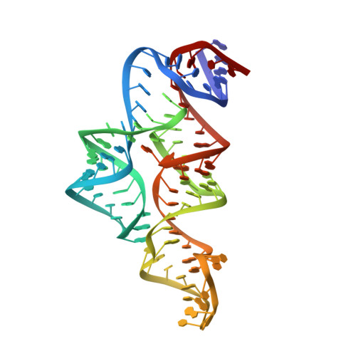
    Cyclic dinucleotides are second messengers that target the adaptor STING and stimulate the innate immune response in mammals. Besides protein receptors, there are bacterial riboswitches that selectively recognize cyclic dinucleotides. We recently discovered a natural riboswitch that targets 3',3'-cGAMP, which is distinguished from the endogenous mammalian signal 2',3'-cGAMP by its backbone connectivity. Here, we report on structures of the aptamer domain of the 3',3'-cGAMP riboswitch from Geobacter in the 3',3'-cGAMP and c-di-GMP bound states. The riboswitch adopts a tuning fork-like architecture with a junctional ligand-binding pocket and different orientations of the arms are correlated with the identity of the bound cyclic dinucleotide. Subsequent biochemical experiments revealed that specificity of ligand recognition can be affected by point mutations outside of the binding pocket, which has implications for both the assignment and reengineering of riboswitches in this structural class.


  • Organizational Affiliation
    • Structural Biology Program, Memorial Sloan-Kettering Cancer Center, New York, NY 10065, USA.

Macromolecule Content 

  • Total Structure Weight: 40.85 kDa 
  • Atom Count: 2,761 
  • Modeled Residue Count: 183 
  • Deposited Residue Count: 183 
  • Unique protein chains: 1
  • Unique nucleic acid chains: 1

Macromolecules


Find similar proteins by:|  3D Structure
Entity ID: 2
MoleculeChains  Sequence LengthOrganismDetailsImage
U1 small nuclear ribonucleoprotein AB [auth P]92Homo sapiensMutation(s): 0 
Gene Names: SNRPA
UniProt & NIH Common Fund Data Resources
Find proteins for P09012 (Homo sapiens)
Explore P09012 
Go to UniProtKB:  P09012
PHAROS:  P09012
GTEx:  ENSG00000077312 
Entity Groups
Sequence Clusters30% Identity50% Identity70% Identity90% Identity95% Identity100% Identity
UniProt GroupP09012
Sequence Annotations
Expand
Reference Sequence
Find similar nucleic acids by:  (by identity cutoff) 
Entity ID: 1
MoleculeChains LengthOrganismImage
RNA (91-MER)A [auth R]91Vibrio cholerae
Sequence Annotations
Expand
Reference Sequence

Small Molecules

Ligands 2 Unique
IDChains Name / Formula / InChI Key2D Diagram3D Interactions
4BW

Query on 4BW


Download:Ideal Coordinates CCD File
D [auth R]2-amino-9-[(2R,3R,3aS,5R,7aR,9R,10R,10aS,12R,14aR)-9-(6-amino-9H-purin-9-yl)-3,5,10,12-tetrahydroxy-5,12-dioxidooctahydro-2H,7H-difuro[3,2-d:3',2'-j][1,3,7,9,2,8]tetraoxadiphosphacyclododecin-2-yl]-1,9-dihydro-6H-purin-6-one
C20 H24 N10 O13 P2
RFCBNSCSPXMEBK-INFSMZHSSA-N
MG

Query on MG


Download:Ideal Coordinates CCD File
C [auth R]MAGNESIUM ION
Mg
JLVVSXFLKOJNIY-UHFFFAOYSA-N

Experimental Data & Validation

Experimental Data

  • Method: X-RAY DIFFRACTION
  • Resolution: 2.08 Å
  • R-Value Free:  0.298 (Depositor), 0.311 (DCC) 
  • R-Value Work:  0.218 (Depositor), 0.235 (DCC) 
  • R-Value Observed: 0.222 (Depositor) 
Space Group: P 1 21 1
Unit Cell:
Length ( Å )Angle ( ˚ )
a = 51.738α = 90
b = 46.836β = 90.44
c = 80.791γ = 90
Software Package:
Software NamePurpose
PHENIXrefinement
XDSdata reduction
XDSdata scaling
PHASERphasing

Structure Validation

View Full Validation Report


Ligand Structure Quality Assessment 


Entry History 

Deposition Data

Revision History  (Full details and data files)

  • Version 1.0: 2015-04-15
    Type: Initial release
  • Version 1.1: 2015-04-22
    Changes: Database references
  • Version 1.2: 2015-07-22
    Changes: Non-polymer description
  • Version 1.3: 2024-02-28
    Changes: Data collection, Database references, Derived calculations, Source and taxonomy, Structure summary