4Y87 | pdb_00004y87

Crystal structure of phosphodiesterase 9 in complex with (R)-C33 (6-{[(1R)-1-(4-chlorophenyl)ethyl]amino}-1-cyclopentyl-1,5-dihydro-4H-pyrazolo[3,4-d]pyrimidin-4-one)


Experimental Data Snapshot

  • Method: X-RAY DIFFRACTION
  • Resolution: 3.10 Å
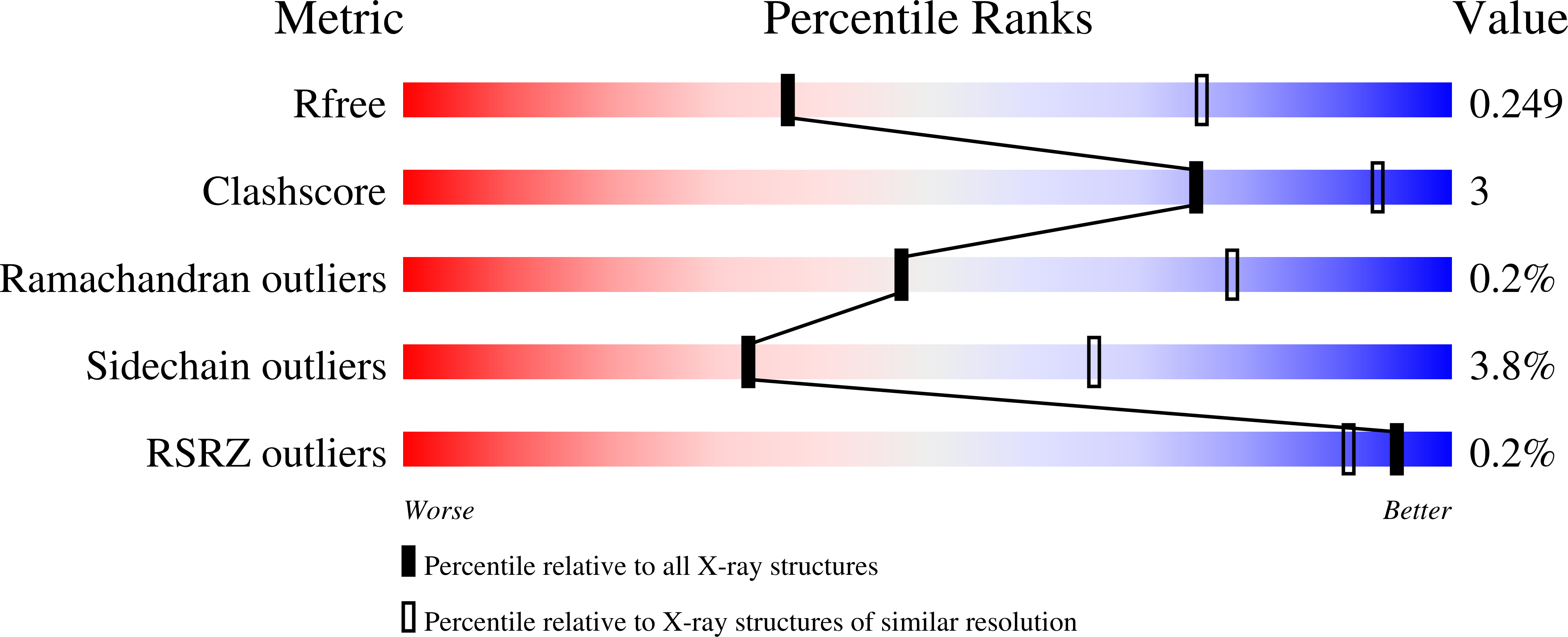
  • R-Value Free: 
    0.247 (Depositor), 0.250 (DCC) 
  • R-Value Work: 
    0.205 (Depositor), 0.210 (DCC) 
  • R-Value Observed: 
    0.207 (Depositor) 

wwPDB Validation   3D Report Full Report


Ligand Structure Quality Assessment 

Created with Raphaël 2.3.0Worse 01 BetterLigand structure goodness of fit to experimental dataBest fitted 49EClick on this verticalbar to view details

This is version 1.4 of the entry. See complete history


Literature

Structural Asymmetry of Phosphodiesterase-9A and a Unique Pocket for Selective Binding of a Potent Enantiomeric Inhibitor.

Huang, M.Shao, Y.Hou, J.Cui, W.Liang, B.Huang, Y.Li, Z.Wu, Y.Zhu, X.Liu, P.Wan, Y.Ke, H.Luo, H.B.

(2015) Mol Pharmacol 88: 836-845

  • DOI: https://doi.org/10.1124/mol.115.099747
  • Primary Citation of Related Structures:  
    4Y86, 4Y87, 4Y8C

  • PubMed Abstract: 

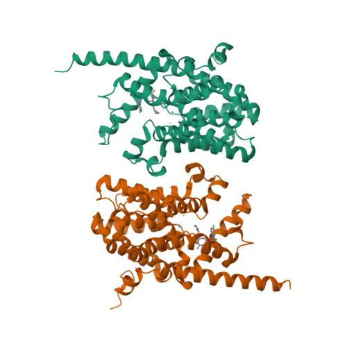
    Phosphodiesterase-9 (PDE9) inhibitors have been studied as potential therapeutics for treatment of central nervous system diseases and diabetes. Here, we report the discovery of a new category of PDE9 inhibitors by rational design on the basis of the crystal structures. The best compound, (S)-6-((1-(4-chlorophenyl)ethyl)amino)-1-cyclopentyl-1,5,6,7-tetrahydro-4H-pyrazolo[3,4-day]pyrimidin-4-one [(S)-C33], has an IC50 value of 11 nM against PDE9 and the racemic C33 has bioavailability of 56.5% in the rat pharmacokinetic model. The crystal structures of PDE9 in the complex with racemic C33, (R)-C33, and (S)-C33 reveal subtle conformational asymmetry of two M-loops in the PDE9 dimer and different conformations of two C33 enantiomers. The structures also identified a small hydrophobic pocket that interacts with the tyrosyl tail of (S)-C33 but not with (R)-C33, and is thus possibly useful for improvement of selectivity of PDE9 inhibitors. The asymmetry of the M-loop and the different interactions of the C33 enantiomers imply the necessity to consider the whole PDE9 dimer in the design of inhibitors.


  • Organizational Affiliation

    School of Chemistry and Chemical Engineering (M.H., J.H., X.Z. Yiq.W.), School of Pharmaceutical Sciences (Y.S., Z.L., Yin.W., P.L., H.-B.L.), Sun Yat-Sen University, Guangzhou, PR China; and Department of Biochemistry and Biophysics and Lineberger Comprehensive Cancer Center, University of North Carolina, Chapel Hill, North Carolina (W.C., B.L., Y.H., H.K.).


Macromolecules
Find similar proteins by:  (by identity cutoff)  |  3D Structure
Entity ID: 1
MoleculeChains Sequence LengthOrganismDetailsImage
High affinity cGMP-specific 3',5'-cyclic phosphodiesterase 9A
A, B
533Homo sapiensMutation(s): 0 
Gene Names: PDE9A
EC: 3.1.4.35
UniProt & NIH Common Fund Data Resources
Find proteins for O76083 (Homo sapiens)
Explore O76083 
Go to UniProtKB:  O76083
PHAROS:  O76083
GTEx:  ENSG00000160191 
Entity Groups  
Sequence Clusters30% Identity50% Identity70% Identity90% Identity95% Identity100% Identity
UniProt GroupO76083
Sequence Annotations
Expand
  • Reference Sequence
Small Molecules
Ligands 3 Unique
IDChains Name / Formula / InChI Key2D Diagram3D Interactions
49E
Query on 49E

Download Ideal Coordinates CCD File 
C [auth A],
F [auth B]
6-{[(1R)-1-(4-chlorophenyl)ethyl]amino}-1-cyclopentyl-1,5-dihydro-4H-pyrazolo[3,4-d]pyrimidin-4-one
C18 H20 Cl N5 O
FIUCLBJMUGCQTF-LLVKDONJSA-N
ZN
Query on ZN

Download Ideal Coordinates CCD File 
D [auth A],
G [auth B]
ZINC ION
Zn
PTFCDOFLOPIGGS-UHFFFAOYSA-N
MG
Query on MG

Download Ideal Coordinates CCD File 
E [auth A],
H [auth B]
MAGNESIUM ION
Mg
JLVVSXFLKOJNIY-UHFFFAOYSA-N
Experimental Data & Validation

Experimental Data

  • Method: X-RAY DIFFRACTION
  • Resolution: 3.10 Å
  • R-Value Free:  0.247 (Depositor), 0.250 (DCC) 
  • R-Value Work:  0.205 (Depositor), 0.210 (DCC) 
  • R-Value Observed: 0.207 (Depositor) 
Space Group: P 41 21 2
Unit Cell:
Length ( Å )Angle ( ˚ )
a = 105.436α = 90
b = 105.436β = 90
c = 270.056γ = 90
Software Package:
Software NamePurpose
REFMACrefinement
HKL-2000data reduction
HKL-2000data scaling
PHASERphasing

Structure Validation

View Full Validation Report


Ligand Structure Quality Assessment 

Created with Raphaël 2.3.0Worse 01 BetterLigand structure goodness of fit to experimental dataBest fitted 49EClick on this verticalbar to view details

Entry History & Funding Information

Deposition Data

  • Released Date: 2015-09-16 
  • Deposition Author(s): Huang, M.

Funding OrganizationLocationGrant Number
National Institutes of Health/National Institute of General Medical Sciences (NIH/NIGMS)United StatesGM59791

Revision History  (Full details and data files)

  • Version 1.0: 2015-09-16
    Type: Initial release
  • Version 1.1: 2015-10-14
    Changes: Database references
  • Version 1.2: 2017-09-20
    Changes: Author supporting evidence, Database references, Derived calculations
  • Version 1.3: 2019-12-25
    Changes: Author supporting evidence
  • Version 1.4: 2024-02-28
    Changes: Data collection, Database references, Derived calculations