4Y5X | pdb_00004y5x

Diabody 305 complex with EpoR


Experimental Data Snapshot

  • Method: X-RAY DIFFRACTION
  • Resolution: 3.15 Å
  • R-Value Free: 
    0.241 (Depositor), 0.240 (DCC) 
  • R-Value Work: 
    0.201 (Depositor), 0.203 (DCC) 
  • R-Value Observed: 
    0.203 (Depositor) 

wwPDB Validation 3D Report Full Report

Validation slider image for 4Y5X

This is version 1.3 of the entry. See complete history. 

Literature

Tuning Cytokine Receptor Signaling by Re-orienting Dimer Geometry with Surrogate Ligands.

Moraga, I., Wernig, G., Wilmes, S., Gryshkova, V., Richter, C.P., Hong, W.J., Sinha, R., Guo, F., Fabionar, H., Wehrman, T.S., Krutzik, P., Demharter, S., Plo, I., Weissman, I.L., Minary, P., Majeti, R., Constantinescu, S.N., Piehler, J., Garcia, K.C.

(2015) Cell 160: 1196-1208

  • DOI: https://doi.org/10.1016/j.cell.2015.02.011
  • Primary Citation Related Structures: 
    4Y5V, 4Y5X, 4Y5Y

  • PubMed Abstract: 

    Most cell-surface receptors for cytokines and growth factors signal as dimers, but it is unclear whether remodeling receptor dimer topology is a viable strategy to "tune" signaling output. We utilized diabodies (DA) as surrogate ligands in a prototypical dimeric receptor-ligand system, the cytokine Erythropoietin (EPO) and its receptor (EpoR), to dimerize EpoR ectodomains in non-native architectures. Diabody-induced signaling amplitudes varied from full to minimal agonism, and structures of these DA/EpoR complexes differed in EpoR dimer orientation and proximity. Diabodies also elicited biased or differential activation of signaling pathways and gene expression profiles compared to EPO. Non-signaling diabodies inhibited proliferation of erythroid precursors from patients with a myeloproliferative neoplasm due to a constitutively active JAK2V617F mutation. Thus, intracellular oncogenic mutations causing ligand-independent receptor activation can be counteracted by extracellular ligands that re-orient receptors into inactive dimer topologies. This approach has broad applications for tuning signaling output for many dimeric receptor systems.


  • Organizational Affiliation: 
    • Howard Hughes Medical Institute, Stanford University School of Medicine, Stanford, CA 94305-5345, USA; Department of Molecular and Cellular Physiology, Stanford University School of Medicine, Stanford, CA 94305-5345, USA.

Macromolecule Content 

  • Total Structure Weight: 207.05 kDa 
  • Atom Count: 13,334 
  • Modeled Residue Count: 1,762 
  • Deposited Residue Count: 1,924 
  • Unique protein chains: 3

Macromolecules

Find similar proteins by:|  3D Structure
Entity ID: 1
MoleculeChains  Sequence LengthOrganismDetailsImage
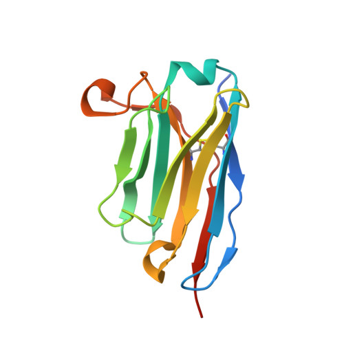
diabody 310 VL domain
A, D, G, J
136Homo sapiensMutation(s): 0 
Entity Groups
Sequence Clusters30% Identity50% Identity70% Identity90% Identity95% Identity100% Identity
Sequence Annotations
Expand
Reference Sequence
Find similar proteins by:|  3D Structure
Entity ID: 2
MoleculeChains  Sequence LengthOrganismDetailsImage
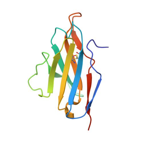
diabody 310 VH domain
B, E, H, K
116Homo sapiensMutation(s): 0 
UniProt
Find proteins for Q5NV67 (Homo sapiens)
Explore Q5NV67 
Go to UniProtKB:  Q5NV67
Entity Groups
Sequence Clusters30% Identity50% Identity70% Identity90% Identity95% Identity100% Identity
UniProt GroupQ5NV67
Sequence Annotations
Expand
Reference Sequence
Find similar proteins by:|  3D Structure
Entity ID: 3
MoleculeChains  Sequence LengthOrganismDetailsImage
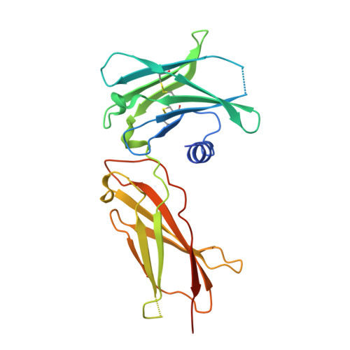
Erythropoietin receptor
C, F, I, L
229Homo sapiensMutation(s): 2 
Gene Names: EPOR
UniProt & NIH Common Fund Data Resources
Find proteins for P19235 (Homo sapiens)
Explore P19235 
Go to UniProtKB:  P19235
PHAROS:  P19235
GTEx:  ENSG00000187266 
Entity Groups
Sequence Clusters30% Identity50% Identity70% Identity90% Identity95% Identity100% Identity
UniProt GroupP19235
Sequence Annotations
Expand
Reference Sequence

Small Molecules

Ligands 2 Unique
IDChains Name / Formula / InChI Key2D Diagram3D Interactions
FLC

Query on FLC


Download:Ideal Coordinates CCD File
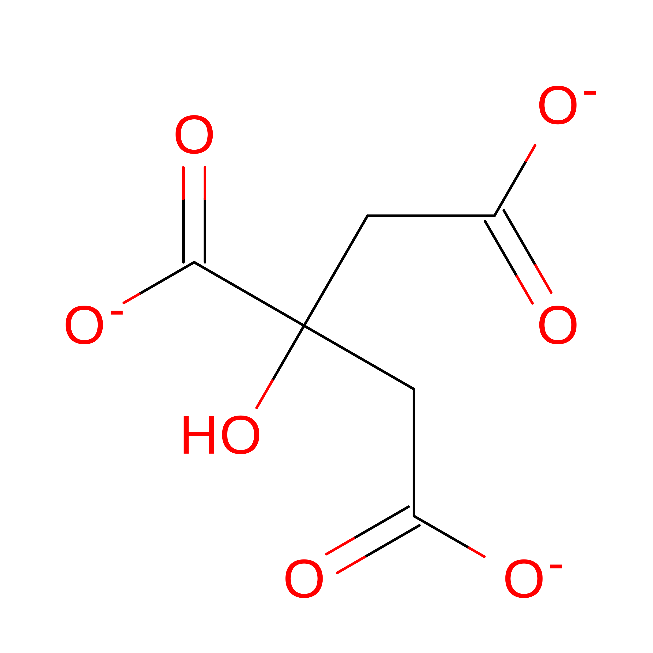
AA [auth K]CITRATE ANION
C6 H5 O7
KRKNYBCHXYNGOX-UHFFFAOYSA-K
PEG

Query on PEG


Download:Ideal Coordinates CCD File
BA [auth L]
CA [auth L]
DA [auth L]
M [auth A]
N [auth B]
BA [auth L],
CA [auth L],
DA [auth L],
M [auth A],
N [auth B],
O [auth B],
P [auth B],
Q [auth C],
R [auth C],
S [auth C],
T [auth E],
U [auth F],
V [auth G],
W [auth G],
X [auth H],
Y [auth J],
Z [auth K]
DI(HYDROXYETHYL)ETHER
C4 H10 O3
MTHSVFCYNBDYFN-UHFFFAOYSA-N

Experimental Data & Validation

Experimental Data

  • Method: X-RAY DIFFRACTION
  • Resolution: 3.15 Å
  • R-Value Free:  0.241 (Depositor), 0.240 (DCC) 
  • R-Value Work:  0.201 (Depositor), 0.203 (DCC) 
  • R-Value Observed: 0.203 (Depositor) 
Space Group: P 41 21 2
Unit Cell:
Length ( Å )Angle ( ˚ )
a = 239.54α = 90
b = 239.54β = 90
c = 132.569γ = 90
Software Package:
Software NamePurpose
PHENIXrefinement
Aimlessdata scaling
PDB_EXTRACTdata extraction
PHASERphasing

Structure Validation

View Full Validation Report


Entry History 

Deposition Data

Revision History  (Full details and data files)

  • Version 1.0: 2015-03-18
    Type: Initial release
  • Version 1.1: 2015-03-25
    Changes: Database references
  • Version 1.2: 2017-11-22
    Changes: Derived calculations, Refinement description, Source and taxonomy, Structure summary
  • Version 1.3: 2024-11-06
    Changes: Data collection, Database references, Structure summary