4Y3U | pdb_00004y3u

The structure of phospholamban bound to the calcium pump SERCA1a


Experimental Data Snapshot

  • Method: X-RAY DIFFRACTION
  • Resolution: 3.51 Å
  • R-Value Free: 
    0.277 (Depositor), 0.271 (DCC) 
  • R-Value Work: 
    0.235 (Depositor), 0.231 (DCC) 
  • R-Value Observed: 
    0.237 (Depositor) 

Starting Model: experimental
View more details

wwPDB Validation 3D Report Full Report

Validation slider image for 4Y3U

This is version 1.4 of the entry. See complete history

Literature

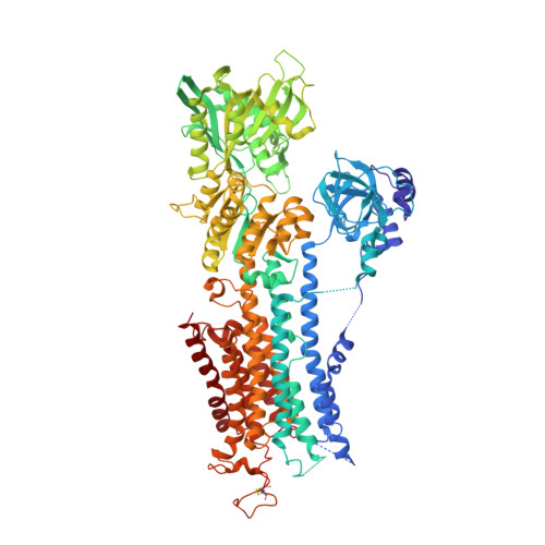
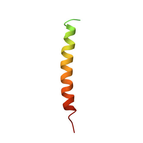
The structural basis for phospholamban inhibition of the calcium pump in sarcoplasmic reticulum.

Akin, B.L.Hurley, T.D.Chen, Z.Jones, L.R.

(2013) J Biological Chem 288: 30181-30191

  • DOI: https://doi.org/10.1074/jbc.M113.501585
  • Primary Citation Related Structures: 
    4KYT, 4Y3U

  • PubMed Abstract: 

    P-type ATPases are a large family of enzymes that actively transport ions across biological membranes by interconverting between high (E1) and low (E2) ion-affinity states; these transmembrane transporters carry out critical processes in nearly all forms of life. In striated muscle, the archetype P-type ATPase, SERCA (sarco(endo)plasmic reticulum Ca(2+)-ATPase), pumps contractile-dependent Ca(2+) ions into the lumen of sarcoplasmic reticulum, which initiates myocyte relaxation and refills the sarcoplasmic reticulum in preparation for the next contraction. In cardiac muscle, SERCA is regulated by phospholamban (PLB), a small inhibitory phosphoprotein that decreases the Ca(2+) affinity of SERCA and attenuates contractile strength. cAMP-dependent phosphorylation of PLB reverses Ca(2+)-ATPase inhibition with powerful contractile effects. Here we present the long sought crystal structure of the PLB-SERCA complex at 2.8-Å resolution. The structure was solved in the absence of Ca(2+) in a novel detergent system employing alkyl mannosides. The structure shows PLB bound to a previously undescribed conformation of SERCA in which the Ca(2+) binding sites are collapsed and devoid of divalent cations (E2-PLB). This new structure represents one of the key unsolved conformational states of SERCA and provides a structural explanation for how dephosphorylated PLB decreases Ca(2+) affinity and depresses cardiac contractility.


  • Organizational Affiliation
    • From the Krannert Institute of Cardiology and the Departments of Medicine and. Electronic address: bbudryns@iu.edu.

Macromolecule Content 

  • Total Structure Weight: 117.48 kDa 
  • Atom Count: 7,812 
  • Modeled Residue Count: 1,016 
  • Deposited Residue Count: 1,067 
  • Unique protein chains: 3

Macromolecules

Find similar proteins by:|  3D Structure
Entity ID: 1
MoleculeChains  Sequence LengthOrganismDetailsImage
Sarcoplasmic/endoplasmic reticulum calcium ATPase 1994Oryctolagus cuniculusMutation(s): 0 
EC: 3.6.3.8 (PDB Primary Data), 7.2.2.10 (UniProt)
Membrane Entity: Yes 
UniProt
Find proteins for P04191 (Oryctolagus cuniculus)
Explore P04191 
Go to UniProtKB:  P04191
Entity Groups
Sequence Clusters30% Identity50% Identity70% Identity90% Identity95% Identity100% Identity
UniProt GroupP04191
Sequence Annotations
Expand
Reference Sequence
Find similar proteins by:|  3D Structure
Entity ID: 2
MoleculeChains  Sequence LengthOrganismDetailsImage
Cardiac phospholamban50Canis lupus familiarisMutation(s): 0 
Gene Names: PLN
Membrane Entity: Yes 
UniProt
Find proteins for P61012 (Canis lupus familiaris)
Explore P61012 
Go to UniProtKB:  P61012
Entity Groups
Sequence Clusters30% Identity50% Identity70% Identity90% Identity95% Identity100% Identity
UniProt GroupP61012
Sequence Annotations
Expand
Reference Sequence
Find similar proteins by:  Sequence   |   3D Structure  
Entity ID: 3
MoleculeChains  Sequence LengthOrganismDetailsImage
Cardiac phospholamban23Canis lupus familiarisMutation(s): 0 
Gene Names: PLN

Small Molecules

Ligands 1 Unique
IDChains Name / Formula / InChI Key2D Diagram3D Interactions
K

Query on K


Download:Ideal Coordinates CCD File
D [auth A]POTASSIUM ION
K
NPYPAHLBTDXSSS-UHFFFAOYSA-N

Experimental Data & Validation

Experimental Data

  • Method: X-RAY DIFFRACTION
  • Resolution: 3.51 Å
  • R-Value Free:  0.277 (Depositor), 0.271 (DCC) 
  • R-Value Work:  0.235 (Depositor), 0.231 (DCC) 
  • R-Value Observed: 0.237 (Depositor) 
Space Group: P 21 21 21
Unit Cell:
Length ( Å )Angle ( ˚ )
a = 61.745α = 90
b = 91.825β = 90
c = 316.256γ = 90
Software Package:
Software NamePurpose
REFMACrefinement
HKL-3000data reduction
HKL-3000data scaling
MOLREPphasing

Structure Validation

View Full Validation Report


Entry History 

& Funding Information

Deposition Data

  • Released Date: 2015-02-25 
  • Deposition Author(s): Hurley, T.D.

Funding OrganizationLocationGrant Number
National Institutes of Health/National Heart, Lung, and Blood Institute (NIH/NHLBI)United StatesR37-HL049428

Revision History  (Full details and data files)

  • Version 1.0: 2015-02-25
    Type: Initial release
  • Version 1.1: 2017-09-20
    Changes: Author supporting evidence, Derived calculations, Source and taxonomy
  • Version 1.2: 2019-12-04
    Changes: Author supporting evidence
  • Version 1.3: 2023-09-27
    Changes: Data collection, Database references, Derived calculations, Refinement description
  • Version 1.4: 2024-11-13
    Changes: Structure summary