4Y3J | pdb_00004y3j

Endothiapepsin in complex with fragment B30


Experimental Data Snapshot

  • Method: X-RAY DIFFRACTION
  • Resolution: 1.31 Å
  • R-Value Free: 
    0.158 (Depositor), 0.158 (DCC) 
  • R-Value Work: 
    0.128 (Depositor), 0.130 (DCC) 
  • R-Value Observed: 
    0.129 (Depositor) 

wwPDB Validation 3D Report Full Report

Validation slider image for 4Y3J

Ligand Structure Quality Assessment 


This is version 1.3 of the entry. See complete history

Literature

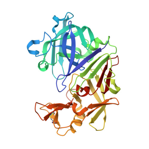
Structures of endothiapepsin-fragment complexes from crystallographic fragment screening using a novel, diverse and affordable 96-compound fragment library.

Huschmann, F.U.Linnik, J.Sparta, K.Uhlein, M.Wang, X.Metz, A.Schiebel, J.Heine, A.Klebe, G.Weiss, M.S.Mueller, U.

(2016) Acta Crystallogr F Struct Biol Commun 72: 346-355

  • DOI: https://doi.org/10.1107/S2053230X16004623
  • Primary Citation Related Structures: 
    4Y38, 4Y3J, 4Y3Y, 4Y48, 4Y4D, 4Y4G, 4Y4J, 4ZE6, 4ZEA

  • PubMed Abstract: 

    Crystallographic screening of the binding of small organic compounds (termed fragments) to proteins is increasingly important for medicinal chemistry-oriented drug discovery. To enable such experiments in a widespread manner, an affordable 96-compound library has been assembled for fragment screening in both academia and industry. The library is selected from already existing protein-ligand structures and is characterized by a broad ligand diversity, including buffer ingredients, carbohydrates, nucleotides, amino acids, peptide-like fragments and various drug-like organic compounds. When applied to the model protease endothiapepsin in a crystallographic screening experiment, a hit rate of nearly 10% was obtained. In comparison to other fragment libraries and considering that no pre-screening was performed, this hit rate is remarkably high. This demonstrates the general suitability of the selected compounds for an initial fragment-screening campaign. The library composition, experimental considerations and time requirements for a complete crystallographic fragment-screening campaign are discussed as well as the nine fully refined obtained endothiapepsin-fragment structures. While most of the fragments bind close to the catalytic centre of endothiapepsin in poses that have been observed previously, two fragments address new sites on the protein surface. ITC measurements show that the fragments bind to endothiapepsin with millimolar affinity.


  • Organizational Affiliation
    • Macromolecular Crystallography, Helmholtz-Zentrum Berlin, Albert-Einstein-Strasse 15, D-12489 Berlin, Germany.

Macromolecule Content 

  • Total Structure Weight: 35.05 kDa 
  • Atom Count: 2,763 
  • Modeled Residue Count: 330 
  • Deposited Residue Count: 330 
  • Unique protein chains: 1

Macromolecules

Find similar proteins by:|  3D Structure
Entity ID: 1
MoleculeChains  Sequence LengthOrganismDetailsImage
Endothiapepsin330Cryphonectria parasiticaMutation(s): 0 
EC: 3.4.23.22
UniProt
Find proteins for P11838 (Cryphonectria parasitica)
Explore P11838 
Go to UniProtKB:  P11838
Entity Groups
Sequence Clusters30% Identity50% Identity70% Identity90% Identity95% Identity100% Identity
UniProt GroupP11838
Sequence Annotations
Expand
Reference Sequence

Small Molecules

Ligands 4 Unique
IDChains Name / Formula / InChI Key2D Diagram3D Interactions
HIC

Query on HIC


Download:Ideal Coordinates CCD File
P [auth A]4-METHYL-HISTIDINE
C7 H11 N3 O2
BRMWTNUJHUMWMS-LURJTMIESA-N
GOL

Query on GOL


Download:Ideal Coordinates CCD File
B [auth A],
C [auth A],
D [auth A],
E [auth A],
F [auth A]
GLYCEROL
C3 H8 O3
PEDCQBHIVMGVHV-UHFFFAOYSA-N
DMS

Query on DMS


Download:Ideal Coordinates CCD File
L [auth A],
M [auth A],
N [auth A],
O [auth A]
DIMETHYL SULFOXIDE
C2 H6 O S
IAZDPXIOMUYVGZ-UHFFFAOYSA-N
ACT

Query on ACT


Download:Ideal Coordinates CCD File
G [auth A],
H [auth A],
I [auth A],
J [auth A],
K [auth A]
ACETATE ION
C2 H3 O2
QTBSBXVTEAMEQO-UHFFFAOYSA-M

Experimental Data & Validation

Experimental Data

  • Method: X-RAY DIFFRACTION
  • Resolution: 1.31 Å
  • R-Value Free:  0.158 (Depositor), 0.158 (DCC) 
  • R-Value Work:  0.128 (Depositor), 0.130 (DCC) 
  • R-Value Observed: 0.129 (Depositor) 
Space Group: P 1 21 1
Unit Cell:
Length ( Å )Angle ( ˚ )
a = 45.216α = 90
b = 73.272β = 109.08
c = 52.787γ = 90
Software Package:
Software NamePurpose
PHENIXrefinement
Cootmodel building
XDSdata reduction
XDSdata scaling

Structure Validation

View Full Validation Report


Ligand Structure Quality Assessment 


Entry History 

& Funding Information

Deposition Data


Funding OrganizationLocationGrant Number
BMBFGermany5116

Revision History  (Full details and data files)

  • Version 1.0: 2016-02-17
    Type: Initial release
  • Version 1.1: 2016-05-04
    Changes: Database references
  • Version 1.2: 2016-05-11
    Changes: Database references
  • Version 1.3: 2025-04-09
    Changes: Data collection, Database references, Structure summary