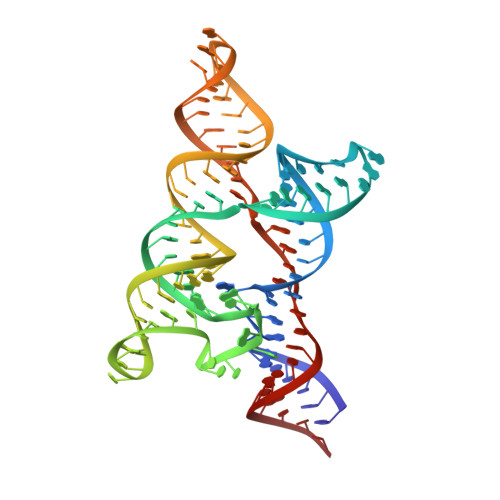
4Y1M

An Escherichia coli yybP-ykoY Mn riboswitch in the Mn2+-free state

  • Classification: RNA
  • Organism(s): Escherichia coli
  • Mutation(s): No 

  • Deposited: 2015-02-08 Released: 2015-04-08 
  • Deposition Author(s): Price, I.R., Ke, A.
  • Funding Organization(s): National Institutes of Health/National Institute of General Medical Sciences (NIH/NIGMS)

Experimental Data Snapshot

  • Method: X-RAY DIFFRACTION
  • Resolution: 3.00 Å
  • R-Value Free: 0.252 
  • R-Value Work: 0.217 
  • R-Value Observed: 0.219 

wwPDB Validation   3D Report Full Report


Ligand Structure Quality Assessment 


This is version 1.4 of the entry. See complete history


Literature

Mn(2+)-Sensing Mechanisms of yybP-ykoY Orphan Riboswitches.

Price, I.R.Gaballa, A.Ding, F.Helmann, J.D.Ke, A.

(2015) Mol Cell 57: 1110-1123

  • DOI: https://doi.org/10.1016/j.molcel.2015.02.016
  • Primary Citation of Related Structures:  
    4Y1I, 4Y1J, 4Y1M

  • PubMed Abstract: 

    Gene regulation in cis by riboswitches is prevalent in bacteria. The yybP-ykoY riboswitch family is quite widespread, yet its ligand and function remained unknown. Here, we characterize the Lactococcus lactis yybP-ykoY orphan riboswitch as a Mn(2+)-dependent transcription-ON riboswitch, with a ∼30-40 μM affinity for Mn(2+). We further determined its crystal structure at 2.7 Å to elucidate the metal sensing mechanism. The riboswitch resembles a hairpin, with two coaxially stacked helices tethered by a four-way junction and a tertiary docking interface. The Mn(2+)-sensing region, strategically located at the highly conserved docking interface, has two metal binding sites. Whereas one site tolerates the binding of either Mg(2+) or Mn(2+), the other site strongly prefers Mn(2+) due to a direct contact from the N7 of an invariable adenosine. Mutagenesis and a Mn(2+)-free E. coli yybP-ykoY structure further reveal that Mn(2+) binding is coupled with stabilization of the Mn(2+)-sensing region and the aptamer domain.


  • Organizational Affiliation

    Department of Molecular Biology and Genetics, Cornell University, 253 Biotechnology Building, Ithaca, NY 14853, USA.


Macromolecules
Find similar nucleic acids by:  (by identity cutoff)  |  3D Structure
Entity ID: 1
MoleculeChains LengthOrganismImage
E. coli yybP-ykoY riboswitchA [auth B],
B [auth A]
107Escherichia coli
Sequence Annotations
Expand
  • Reference Sequence
Small Molecules
Ligands 3 Unique
IDChains Name / Formula / InChI Key2D Diagram3D Interactions
GTP
Query on GTP

Download Ideal Coordinates CCD File 
GB [auth A],
N [auth B]
GUANOSINE-5'-TRIPHOSPHATE
C10 H16 N5 O14 P3
XKMLYUALXHKNFT-UUOKFMHZSA-N
SR
Query on SR

Download Ideal Coordinates CCD File 
AA [auth B]
AB [auth A]
BA [auth B]
BB [auth A]
C [auth B]
AA [auth B],
AB [auth A],
BA [auth B],
BB [auth A],
C [auth B],
CA [auth B],
CB [auth A],
D [auth B],
DA [auth B],
DB [auth A],
E [auth B],
EA [auth B],
EB [auth A],
F [auth B],
FA [auth B],
FB [auth A],
G [auth B],
GA [auth B],
H [auth B],
HA [auth B],
HB [auth A],
I [auth B],
IA [auth B],
IB [auth A],
J [auth B],
JA [auth B],
JB [auth A],
K [auth B],
KA [auth B],
KB [auth A],
L [auth B],
LA [auth B],
LB [auth A],
MA [auth B],
MB [auth A],
NA [auth B],
NB [auth A],
O [auth B],
OA [auth B],
OB [auth A],
P [auth B],
PA [auth B],
PB [auth A],
Q [auth B],
QA [auth B],
QB [auth A],
R [auth B],
RA [auth B],
RB [auth A],
S [auth B],
SA [auth B],
SB [auth A],
T [auth B],
TA [auth B],
TB [auth A],
U [auth B],
UA [auth B],
UB [auth A],
V [auth B],
VB [auth A],
W [auth B],
X [auth B],
Y [auth B],
Z [auth B],
ZA [auth A]
STRONTIUM ION
Sr
PWYYWQHXAPXYMF-UHFFFAOYSA-N
MG
Query on MG

Download Ideal Coordinates CCD File 
M [auth B]
VA [auth B]
WA [auth B]
WB [auth A]
XA [auth B]
M [auth B],
VA [auth B],
WA [auth B],
WB [auth A],
XA [auth B],
XB [auth A],
YA [auth B],
YB [auth A],
ZB [auth A]
MAGNESIUM ION
Mg
JLVVSXFLKOJNIY-UHFFFAOYSA-N
Experimental Data & Validation

Experimental Data

  • Method: X-RAY DIFFRACTION
  • Resolution: 3.00 Å
  • R-Value Free: 0.252 
  • R-Value Work: 0.217 
  • R-Value Observed: 0.219 
  • Space Group: H 3 2
Unit Cell:
Length ( Å )Angle ( ˚ )
a = 159.384α = 90
b = 159.384β = 90
c = 277.108γ = 120
Software Package:
Software NamePurpose
PHENIXrefinement
XDSdata reduction
Aimlessdata scaling
SHELXphasing

Structure Validation

View Full Validation Report


Ligand Structure Quality Assessment 


Entry History & Funding Information

Deposition Data


Funding OrganizationLocationGrant Number
National Institutes of Health/National Institute of General Medical Sciences (NIH/NIGMS)United StatesGM086766
National Institutes of Health/National Institute of General Medical Sciences (NIH/NIGMS)United StatesGM 102543

Revision History  (Full details and data files)

  • Version 1.0: 2015-04-08
    Type: Initial release
  • Version 1.1: 2016-07-20
    Changes: Data collection
  • Version 1.2: 2017-09-20
    Changes: Author supporting evidence, Derived calculations
  • Version 1.3: 2019-12-25
    Changes: Author supporting evidence
  • Version 1.4: 2024-02-28
    Changes: Data collection, Database references, Derived calculations