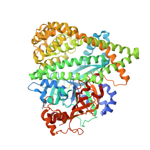
4Y06

Crystal structure of the DAP BII (G675R) dipeptide complex


Experimental Data Snapshot

  • Method: X-RAY DIFFRACTION
  • Resolution: 2.18 Å
  • R-Value Free: 0.235 
  • R-Value Work: 0.176 
  • R-Value Observed: 0.179 

Starting Model: experimental
View more details

wwPDB Validation   3D Report Full Report


This is version 1.3 of the entry. See complete history


Literature

Structural and mutational analyses of dipeptidyl peptidase 11 from Porphyromonas gingivalis reveal the molecular basis for strict substrate specificity.

Sakamoto, Y.Suzuki, Y.Iizuka, I.Tateoka, C.Roppongi, S.Fujimoto, M.Inaka, K.Tanaka, H.Yamada, M.Ohta, K.Gouda, H.Nonaka, T.Ogasawara, W.Tanaka, N.

(2015) Sci Rep 5: 11151-11151

  • DOI: https://doi.org/10.1038/srep11151
  • Primary Citation of Related Structures:  
    4XZY, 4Y01, 4Y02, 4Y04, 4Y06

  • PubMed Abstract: 

    The dipeptidyl peptidase 11 from Porphyromonas gingivalis (PgDPP11) belongs to the S46 family of serine peptidases and preferentially cleaves substrates with Asp/Glu at the P1 position. The molecular mechanism underlying the substrate specificity of PgDPP11, however, is unknown. Here, we report the crystal structure of PgDPP11. The enzyme contains a catalytic domain with a typical double β-barrel fold and a recently identified regulatory α-helical domain. Crystal structure analyses, docking studies, and biochemical studies revealed that the side chain of Arg673 in the S1 subsite is essential for recognition of the Asp/Glu side chain at the P1 position of the bound substrate. Because S46 peptidases are not found in mammals and the Arg673 is conserved among DPP11s, we anticipate that DPP11s could be utilised as targets for antibiotics. In addition, the present structure analyses could be useful templates for the design of specific inhibitors of DPP11s from pathogenic organisms.


  • Organizational Affiliation

    School of Pharmacy, Iwate Medical University, 2-1-1 Nishitokuta, Yahaba, Iwate 028-3694, Japan.


Macromolecules
Find similar proteins by:  (by identity cutoff)  |  3D Structure
Entity ID: 1
MoleculeChains Sequence LengthOrganismDetailsImage
Dipeptidyl aminopeptidase BII
A, B
722Pseudoxanthomonas mexicanaMutation(s): 1 
Gene Names: dapb2
EC: 3.4.14
UniProt
Find proteins for V5YM14 (Pseudoxanthomonas mexicana)
Explore V5YM14 
Go to UniProtKB:  V5YM14
Entity Groups  
Sequence Clusters30% Identity50% Identity70% Identity90% Identity95% Identity100% Identity
UniProt GroupV5YM14
Sequence Annotations
Expand
  • Reference Sequence
Small Molecules
Ligands 4 Unique
IDChains Name / Formula / InChI Key2D Diagram3D Interactions
GLU
Query on GLU

Download Ideal Coordinates CCD File 
D [auth A],
O [auth B]
GLUTAMIC ACID
C5 H9 N O4
WHUUTDBJXJRKMK-VKHMYHEASA-N
LEU
Query on LEU

Download Ideal Coordinates CCD File 
C [auth A],
N [auth B]
LEUCINE
C6 H13 N O2
ROHFNLRQFUQHCH-YFKPBYRVSA-N
GOL
Query on GOL

Download Ideal Coordinates CCD File 
E [auth A]
F [auth A]
G [auth A]
H [auth A]
I [auth A]
E [auth A],
F [auth A],
G [auth A],
H [auth A],
I [auth A],
J [auth A],
P [auth B],
Q [auth B],
R [auth B],
S [auth B],
T [auth B],
U [auth B],
V [auth B],
W [auth B]
GLYCEROL
C3 H8 O3
PEDCQBHIVMGVHV-UHFFFAOYSA-N
ZN
Query on ZN

Download Ideal Coordinates CCD File 
K [auth A],
L [auth A],
M [auth A],
X [auth B],
Y [auth B]
ZINC ION
Zn
PTFCDOFLOPIGGS-UHFFFAOYSA-N
Experimental Data & Validation

Experimental Data

  • Method: X-RAY DIFFRACTION
  • Resolution: 2.18 Å
  • R-Value Free: 0.235 
  • R-Value Work: 0.176 
  • R-Value Observed: 0.179 
  • Space Group: P 43 21 2
Unit Cell:
Length ( Å )Angle ( ˚ )
a = 122.42α = 90
b = 122.42β = 90
c = 222.06γ = 90
Software Package:
Software NamePurpose
REFMACrefinement
XDSdata reduction

Structure Validation

View Full Validation Report


Entry History & Funding Information

Deposition Data


Funding OrganizationLocationGrant Number
Japan Society for the Promotion of ScienceJapan25462872

Revision History  (Full details and data files)

  • Version 1.0: 2015-07-15
    Type: Initial release
  • Version 1.1: 2020-02-05
    Changes: Data collection, Derived calculations, Source and taxonomy
  • Version 1.2: 2023-11-08
    Changes: Data collection, Database references, Derived calculations, Refinement description
  • Version 1.3: 2024-11-06
    Changes: Structure summary