4XZQ

Nucleosome disassembly by RSC and SWI/SNF is enhanced by H3 acetylation near the nucleosome dyad axis


Experimental Data Snapshot

  • Method: X-RAY DIFFRACTION
  • Resolution: 2.40 Å
  • R-Value Free: 0.299 
  • R-Value Work: 0.256 

Starting Model: experimental
View more details

wwPDB Validation   3D Report Full Report


This is version 1.6 of the entry. See complete history


Literature

Histone Acetylation near the Nucleosome Dyad Axis Enhances Nucleosome Disassembly by RSC and SWI/SNF.

Chatterjee, N.North, J.A.Dechassa, M.L.Manohar, M.Prasad, R.Luger, K.Ottesen, J.J.Poirier, M.G.Bartholomew, B.

(2015) Mol Cell Biol 35: 4083-4092

  • DOI: https://doi.org/10.1128/MCB.00441-15
  • Primary Citation of Related Structures:  
    4XZQ, 4YS3, 4Z66

  • PubMed Abstract: 

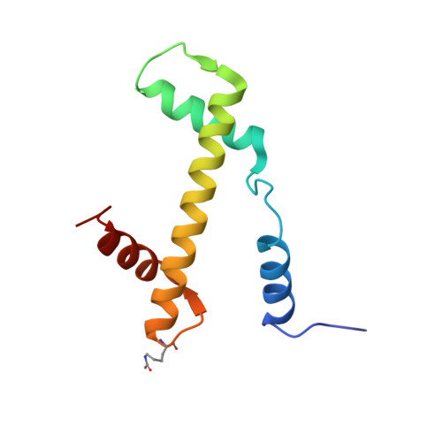
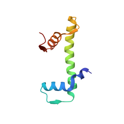
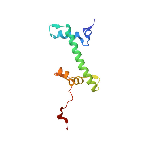
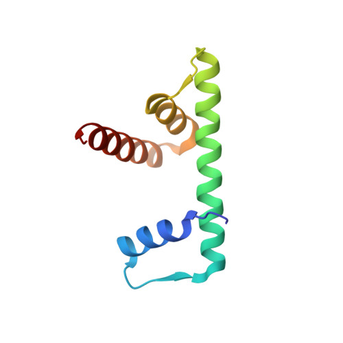
    Signaling associated with transcription activation occurs through posttranslational modification of histones and is best exemplified by lysine acetylation. Lysines are acetylated in histone tails and the core domain/lateral surface of histone octamers. While acetylated lysines in histone tails are frequently recognized by other factors referred to as "readers," which promote transcription, the mechanistic role of the modifications in the lateral surface of the histone octamer remains unclear. By using X-ray crystallography, we found that acetylated lysines 115 and 122 in histone H3 are solvent accessible, but in biochemical assays they appear not to interact with the bromodomains of SWI/SNF and RSC to enhance recruitment or nucleosome mobilization, as previously shown for acetylated lysines in H3 histone tails. Instead, we found that acetylation of lysines 115 and 122 increases the predisposition of nucleosomes for disassembly by SWI/SNF and RSC up to 7-fold, independent of bromodomains, and only in conjunction with contiguous nucleosomes. Thus, in combination with SWI/SNF and RSC, acetylation of lateral surface lysines in the histone octamer serves as a crucial regulator of nucleosomal dynamics distinct from the histone code readers and writers.


  • Organizational Affiliation

    Department of Epigenetics and Molecular Carcinogenesis, UT M. D. Anderson Cancer Center, Smithville, Texas, USA.


Macromolecules

Find similar proteins by:  (by identity cutoff)  |  3D Structure
Entity ID: 1
MoleculeChains Sequence LengthOrganismDetailsImage
Histone H3.2
A, E
98Xenopus laevisMutation(s): 0 
UniProt
Find proteins for P84233 (Xenopus laevis)
Go to UniProtKB:  P84233
Sequence Annotations
Expand
  • Reference Sequence
Find similar proteins by:  (by identity cutoff)  |  3D Structure
Entity ID: 2
MoleculeChains Sequence LengthOrganismDetailsImage
Histone H4
B, F
79Xenopus laevisMutation(s): 0 
UniProt
Find proteins for P62799 (Xenopus laevis)
Go to UniProtKB:  P62799
Sequence Annotations
Expand
  • Reference Sequence
Find similar proteins by:  (by identity cutoff)  |  3D Structure
Entity ID: 3
MoleculeChains Sequence LengthOrganismDetailsImage
Histone H2A
C, G
107Xenopus laevisMutation(s): 0 
Gene Names: hist1h2ajLOC494591
UniProt
Find proteins for P06897 (Xenopus laevis)
Go to UniProtKB:  P06897
Sequence Annotations
Expand
  • Reference Sequence
Find similar proteins by:  (by identity cutoff)  |  3D Structure
Entity ID: 4
MoleculeChains Sequence LengthOrganismDetailsImage
Histone H2B 1.1
D, H
93Xenopus laevisMutation(s): 0 
UniProt
Find proteins for P02281 (Xenopus laevis)
Go to UniProtKB:  P02281
Sequence Annotations
Expand
  • Reference Sequence
Find similar nucleic acids by:  (by identity cutoff)  |  3D Structure
Entity ID: 5
MoleculeChains LengthOrganismImage
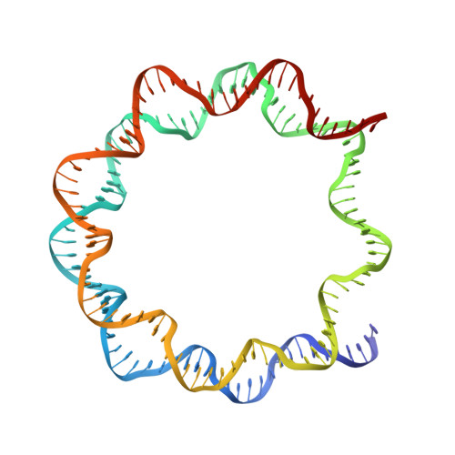
DNA (147-MER)147Homo sapiens
Sequence Annotations
Expand
  • Reference Sequence
Find similar nucleic acids by:  (by identity cutoff)  |  3D Structure
Entity ID: 6
MoleculeChains LengthOrganismImage
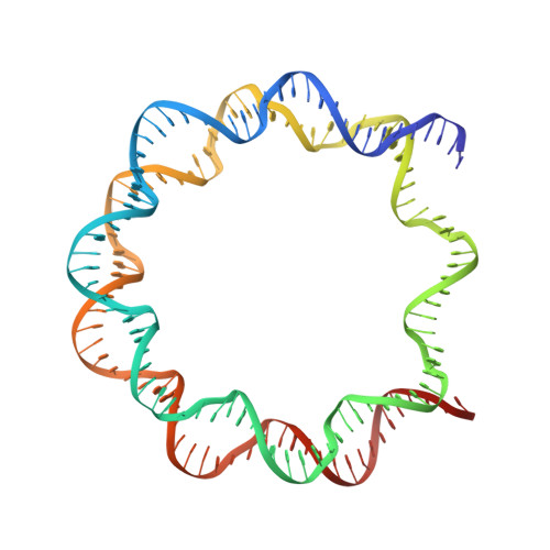
DNA (147-MER)147Homo sapiens
Sequence Annotations
Expand
  • Reference Sequence
Small Molecules
Modified Residues  1 Unique
IDChains TypeFormula2D DiagramParent
ALY
Query on ALY
A, E
L-PEPTIDE LINKINGC8 H16 N2 O3LYS
Experimental Data & Validation

Experimental Data

  • Method: X-RAY DIFFRACTION
  • Resolution: 2.40 Å
  • R-Value Free: 0.299 
  • R-Value Work: 0.256 
  • Space Group: P 21 21 21
Unit Cell:
Length ( Å )Angle ( ˚ )
a = 105.4α = 90
b = 109.57β = 90
c = 180.81γ = 90
Software Package:
Software NamePurpose
CNSrefinement
d*TREKdata reduction
d*TREKdata scaling
Cootmodel building
CNSphasing

Structure Validation

View Full Validation Report


Entry History & Funding Information

Deposition Data


Funding OrganizationLocationGrant Number
National Institutes of Health/National Institute of General Medical Sciences (NIH/NIGMS)United StatesGM088409
National Institutes of Health/National Institute of General Medical Sciences (NIH/NIGMS)United StatesGM048413
National Institutes of Health/National Institute of General Medical Sciences (NIH/NIGMS)United StatesGM083055

Revision History  (Full details and data files)

  • Version 1.0: 2015-10-14
    Type: Initial release
  • Version 1.1: 2015-11-11
    Changes: Database references
  • Version 1.2: 2018-01-17
    Changes: Author supporting evidence, Database references, Derived calculations
  • Version 1.3: 2019-12-25
    Changes: Author supporting evidence
  • Version 1.4: 2022-03-23
    Changes: Author supporting evidence, Database references
  • Version 1.5: 2023-09-27
    Changes: Data collection, Refinement description
  • Version 1.6: 2023-11-15
    Changes: Data collection