4XZ7 | pdb_00004xz7

Crystal structure of a TGase


Experimental Data Snapshot

  • Method: X-RAY DIFFRACTION
  • Resolution: 2.10 Å
  • R-Value Free: 
    0.236 (Depositor), 0.237 (DCC) 
  • R-Value Work: 
    0.192 (Depositor), 0.197 (DCC) 
  • R-Value Observed: 
    0.194 (Depositor) 

wwPDB Validation 3D Report Full Report

Validation slider image for 4XZ7

This is version 1.3 of the entry. See complete history

Literature

Functional and Structural Characterization of the Antiphagocytic Properties of a Novel Transglutaminase from Streptococcus suis

Yu, J.Pian, Y.Ge, J.Guo, J.Zheng, Y.Jiang, H.Hao, H.Yuan, Y.Jiang, Y.Yang, M.

(2015) J Biological Chem 290: 19081-19092

  • DOI: https://doi.org/10.1074/jbc.M115.643338
  • Primary Citation Related Structures: 
    4XZ7

  • PubMed Abstract: 

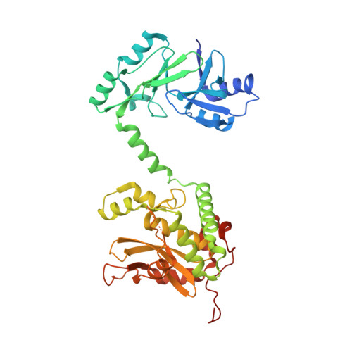
    Streptococcus suis serotype 2 (Ss2) is an important swine and human zoonotic pathogen. In the present study, we identified a novel secreted immunogenic protein, SsTGase, containing a highly conserved eukaryotic-like transglutaminase (TGase) domain at the N terminus. We found that inactivation of SsTGase significantly reduced the virulence of Ss2 in a pig infection model and impaired its antiphagocytosis in human blood. We further solved the crystal structure of the N-terminal portion of the protein in homodimer form at 2.1 Å. Structure-based mutagenesis and biochemical studies suggested that disruption of the homodimer directly resulted in the loss of its TGase activity and antiphagocytic ability. Characterization of SsTGase as a novel virulence factor of Ss2 by acting as a TGase would be beneficial for developing new therapeutic agents against Ss2 infections.


  • Organizational Affiliation
    • From the State Key Laboratory of Pathogen and Biosecurity, Beijing Institute of Microbiology and Epidemiology, Beijing 100071, China, Key Laboratory for Protein Sciences of Ministry of Education, Tsinghua-Peking Center for Life Sciences, School of Life Sciences, Tsinghua University, Beijing 100084, China, and.

Macromolecule Content 

  • Total Structure Weight: 91.47 kDa 
  • Atom Count: 6,515 
  • Modeled Residue Count: 760 
  • Deposited Residue Count: 798 
  • Unique protein chains: 1

Macromolecules

Find similar proteins by:|  3D Structure
Entity ID: 1
MoleculeChains  Sequence LengthOrganismDetailsImage
Putative uncharacterized protein
A, B
399Streptococcus suisMutation(s): 0 
UniProt
Find proteins for A5JSJ3 (Streptococcus suis)
Explore A5JSJ3 
Go to UniProtKB:  A5JSJ3
Entity Groups
Sequence Clusters30% Identity50% Identity70% Identity90% Identity95% Identity100% Identity
UniProt GroupA5JSJ3
Sequence Annotations
Expand
Reference Sequence

Experimental Data & Validation

Experimental Data

  • Method: X-RAY DIFFRACTION
  • Resolution: 2.10 Å
  • R-Value Free:  0.236 (Depositor), 0.237 (DCC) 
  • R-Value Work:  0.192 (Depositor), 0.197 (DCC) 
  • R-Value Observed: 0.194 (Depositor) 
Space Group: P 21 21 21
Unit Cell:
Length ( Å )Angle ( ˚ )
a = 89.133α = 90
b = 91.935β = 90
c = 102.172γ = 90
Software Package:
Software NamePurpose
PHENIXrefinement
HKL-2000data reduction
HKL-2000data scaling
PHASERphasing

Structure Validation

View Full Validation Report


Entry History 

Deposition Data

Revision History  (Full details and data files)

  • Version 1.0: 2015-06-24
    Type: Initial release
  • Version 1.1: 2015-07-01
    Changes: Database references
  • Version 1.2: 2015-08-12
    Changes: Database references
  • Version 1.3: 2024-03-20
    Changes: Data collection, Database references, Derived calculations