4XXG | pdb_00004xxg

Structure of protonated Cholesterol Oxidase from Streptomyces SA-COO


Experimental Data Snapshot

  • Method: X-RAY DIFFRACTION
  • Resolution: 0.85 Å
  • R-Value Free: 
    0.124 (Depositor), 0.120 (DCC) 
  • R-Value Work: 
    0.113 (Depositor), 0.108 (DCC) 
  • R-Value Observed: 
    0.114 (Depositor) 

Starting Model: experimental
View more details

wwPDB Validation 3D Report Full Report

Validation slider image for 4XXG

Ligand Structure Quality Assessment 


This is version 1.1 of the entry. See complete history

Literature

Production and characterization of recombinant perdeuterated cholesterol oxidase.

Golden, E.Attwood, P.V.Duff, A.P.Meilleur, F.Vrielink, A.

(2015) Anal Biochem 485: 102-108

  • DOI: https://doi.org/10.1016/j.ab.2015.06.008
  • Primary Citation Related Structures: 
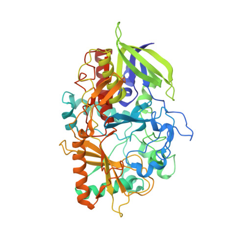
    4XWR, 4XXG

  • PubMed Abstract: 

    Cholesterol oxidase (CO) is a FAD (flavin adenine dinucleotide) containing enzyme that catalyzes the oxidization and isomerization of cholesterol. Studies directed toward elucidating the catalytic mechanism of CO will provide an important general understanding of Flavin-assisted redox catalysis. Hydrogen atoms play an important role in enzyme catalysis; however, they are not readily visualized in protein X-ray diffraction structures. Neutron crystallography is an ideal method for directly visualizing hydrogen positions at moderate resolutions because hydrogen and deuterium have comparable neutron scattering lengths to other heavy atoms present in proteins. The negative coherent and large incoherent scattering lengths of hydrogen atoms in neutron diffraction experiments can be circumvented by replacing hydrogen atoms with its isotope, deuterium. The perdeuterated form of CO was successfully expressed from minimal medium, purified, and crystallized. X-ray crystallographic structures of the enzyme in the perdeuterated and hydrogenated states confirm that there are no apparent structural differences between the two enzyme forms. Kinetic assays demonstrate that perdeuterated and hydrogenated enzymes are functionally identical. Together, structural and functional studies indicate that the perdeuterated protein is suitable for structural studies by neutron crystallography directed at understanding the role of hydrogen atoms in enzyme catalysis.


  • Organizational Affiliation
    • School of Chemistry and Biochemistry, University of Western Australia, Crawley, WA 6009, Australia.

Macromolecule Content 

  • Total Structure Weight: 56.58 kDa 
  • Atom Count: 5,023 
  • Modeled Residue Count: 499 
  • Deposited Residue Count: 510 
  • Unique protein chains: 1

Macromolecules

Find similar proteins by:|  3D Structure
Entity ID: 1
MoleculeChains  Sequence LengthOrganismDetailsImage
Cholesterol oxidase510Streptomyces sp. SA-COOMutation(s): 0 
Gene Names: choA
EC: 1.1.3.6 (PDB Primary Data), 5.3.3.1 (PDB Primary Data)
UniProt
Find proteins for P12676 (Streptomyces sp. (strain SA-COO))
Explore P12676 
Go to UniProtKB:  P12676
Entity Groups
Sequence Clusters30% Identity50% Identity70% Identity90% Identity95% Identity100% Identity
UniProt GroupP12676
Sequence Annotations
Expand
Reference Sequence

Small Molecules

Ligands 1 Unique
IDChains Name / Formula / InChI Key2D Diagram3D Interactions
FAD

Query on FAD


Download:Ideal Coordinates CCD File
B [auth A]FLAVIN-ADENINE DINUCLEOTIDE
C27 H33 N9 O15 P2
VWWQXMAJTJZDQX-UYBVJOGSSA-N

Experimental Data & Validation

Experimental Data

  • Method: X-RAY DIFFRACTION
  • Resolution: 0.85 Å
  • R-Value Free:  0.124 (Depositor), 0.120 (DCC) 
  • R-Value Work:  0.113 (Depositor), 0.108 (DCC) 
  • R-Value Observed: 0.114 (Depositor) 
Space Group: P 1 21 1
Unit Cell:
Length ( Å )Angle ( ˚ )
a = 51.299α = 90
b = 72.761β = 105.13
c = 63.03γ = 90
Software Package:
Software NamePurpose
PHENIXrefinement
PDB_EXTRACTdata extraction
XDSdata reduction
SCALAdata scaling
PHASERphasing

Structure Validation

View Full Validation Report


Ligand Structure Quality Assessment 


Entry History 

Deposition Data

Revision History  (Full details and data files)

  • Version 1.0: 2015-12-30
    Type: Initial release
  • Version 1.1: 2023-09-27
    Changes: Data collection, Database references, Derived calculations, Refinement description