4XX1 | pdb_00004xx1

Low resolution structure of LCAT in complex with Fab1


Experimental Data Snapshot

  • Method: X-RAY DIFFRACTION
  • Resolution: 3.60 Å
  • R-Value Free: 
    0.280 (Depositor), 0.277 (DCC) 
  • R-Value Work: 
    0.196 (Depositor), 0.202 (DCC) 
  • R-Value Observed: 
    0.200 (Depositor) 

Starting Model: experimental
View more details

wwPDB Validation 3D Report Full Report

Validation slider image for 4XX1

Ligand Structure Quality Assessment 


This is version 1.5 of the entry. See complete history

Literature

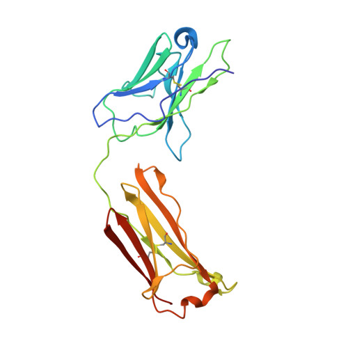
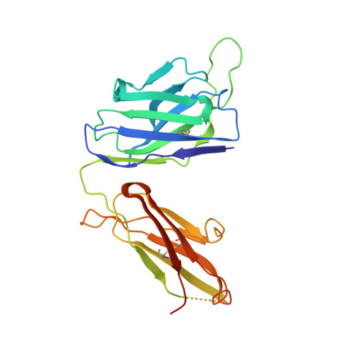
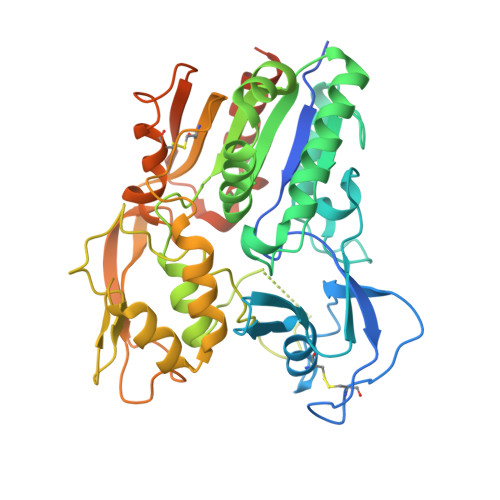
The high-resolution crystal structure of human LCAT.

Piper, D.E.Romanow, W.G.Gunawardane, R.N.Fordstrom, P.Masterman, S.Pan, O.Thibault, S.T.Zhang, R.Meininger, D.Schwarz, M.Wang, Z.King, C.Zhou, M.Walker, N.P.

(2015) J Lipid Res 56: 1711-1719

  • DOI: https://doi.org/10.1194/jlr.M059873
  • Primary Citation Related Structures: 
    4XWG, 4XX1

  • PubMed Abstract: 

    LCAT is intimately involved in HDL maturation and is a key component of the reverse cholesterol transport (RCT) pathway which removes excess cholesterol molecules from the peripheral tissues to the liver for excretion. Patients with loss-of-function LCAT mutations exhibit low levels of HDL cholesterol and corneal opacity. Here we report the 2.65 Å crystal structure of the human LCAT protein. Crystallization required enzymatic removal of N-linked glycans and complex formation with a Fab fragment from a tool antibody. The crystal structure reveals that LCAT has an α/β hydrolase core with two additional subdomains that play important roles in LCAT function. Subdomain 1 contains the region of LCAT shown to be required for interfacial activation, while subdomain 2 contains the lid and amino acids that shape the substrate binding pocket. Mapping the naturally occurring mutations onto the structure provides insight into how they may affect LCAT enzymatic activity.


  • Organizational Affiliation
    • Therapeutic Discovery Amgen Inc., South San Francisco, CA 94080.

Macromolecule Content 

  • Total Structure Weight: 290.93 kDa 
  • Atom Count: 17,233 
  • Modeled Residue Count: 2,186 
  • Deposited Residue Count: 2,619 
  • Unique protein chains: 3

Macromolecules

Find similar proteins by:|  3D Structure
Entity ID: 1
MoleculeChains  Sequence LengthOrganismDetailsImage
Fab1 light chainA [auth L],
D [auth G],
G [auth O]
213Homo sapiensMutation(s): 0 
Find similar proteins by:|  3D Structure
Entity ID: 2
MoleculeChains  Sequence LengthOrganismDetailsImage
Fab1 heavy chainB [auth H],
E,
H [auth M]
238Homo sapiensMutation(s): 0 
Find similar proteins by:|  3D Structure
Entity ID: 3
MoleculeChains  Sequence LengthOrganismDetailsImage
Phosphatidylcholine-sterol acyltransferaseC [auth A],
F [auth B],
I [auth J]
422Homo sapiensMutation(s): 0 
Gene Names: LCAT
EC: 2.3.1.43 (PDB Primary Data), 3.1.1.47 (UniProt)
UniProt & NIH Common Fund Data Resources
Find proteins for P04180 (Homo sapiens)
Explore P04180 
Go to UniProtKB:  P04180
PHAROS:  P04180
GTEx:  ENSG00000213398 
Entity Groups
Sequence Clusters30% Identity50% Identity70% Identity90% Identity95% Identity100% Identity
UniProt GroupP04180
Glycosylation
Glycosylation Sites: 3Go to GlyGen: P04180-1
Sequence Annotations
Expand
Reference Sequence

Small Molecules

Ligands 1 Unique
IDChains Name / Formula / InChI Key2D Diagram3D Interactions
NAG

Query on NAG


Download:Ideal Coordinates CCD File
J [auth A]
K [auth A]
L [auth A]
M [auth B]
N [auth B]
J [auth A],
K [auth A],
L [auth A],
M [auth B],
N [auth B],
O [auth B],
P [auth J],
Q [auth J],
R [auth J]
2-acetamido-2-deoxy-beta-D-glucopyranose
C8 H15 N O6
OVRNDRQMDRJTHS-FMDGEEDCSA-N

Experimental Data & Validation

Experimental Data

  • Method: X-RAY DIFFRACTION
  • Resolution: 3.60 Å
  • R-Value Free:  0.280 (Depositor), 0.277 (DCC) 
  • R-Value Work:  0.196 (Depositor), 0.202 (DCC) 
  • R-Value Observed: 0.200 (Depositor) 
Space Group: P 32
Unit Cell:
Length ( Å )Angle ( ˚ )
a = 166.419α = 90
b = 166.419β = 90
c = 97.66γ = 120
Software Package:
Software NamePurpose
MOSFLMdata reduction
PHENIXrefinement
PDB_EXTRACTdata extraction
SCALAdata scaling
PHASERphasing

Structure Validation

View Full Validation Report


Ligand Structure Quality Assessment 


Entry History 

Deposition Data

Revision History  (Full details and data files)

  • Version 1.0: 2015-07-29
    Type: Initial release
  • Version 1.1: 2015-08-05
    Changes: Database references
  • Version 1.2: 2015-09-09
    Changes: Database references
  • Version 1.3: 2020-07-29
    Type: Remediation
    Reason: Carbohydrate remediation
    Changes: Data collection, Database references, Derived calculations, Structure summary
  • Version 1.4: 2023-09-27
    Changes: Data collection, Database references, Refinement description, Structure summary
  • Version 1.5: 2024-11-06
    Changes: Structure summary