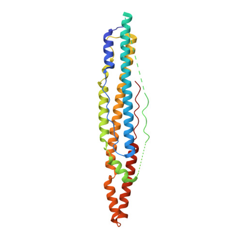
Structure of EspB, a secreted substrate of the ESX-1 secretion system of Mycobacterium tuberculosis.
Korotkova, N., Piton, J., Wagner, J.M., Boy-Rottger, S., Japaridze, A., Evans, T.J., Cole, S.T., Pojer, F., Korotkov, K.V.(2015) J Struct Biol 191: 236-244
- PubMed: 26051906
- DOI: https://doi.org/10.1016/j.jsb.2015.06.003
- Primary Citation of Related Structures:
4XWP, 4XXN, 4XXX, 4XY3 - PubMed Abstract:
Mycobacterium tuberculosis secretes multiple virulence factors during infection via the general Sec and Tat pathways, and via specialized ESX secretion systems, also referred to as type VII secretion systems. The ESX-1 secretion system is an important virulence determinant because deletion of ESX-1 leads to attenuation of M. tuberculosis. ESX-1 secreted protein B (EspB) contains putative PE (Pro-Glu) and PPE (Pro-Pro-Glu) domains, and a C-terminal domain, which is processed by MycP1 protease during secretion. We determined the crystal structure of PE-PPE domains of EspB, which represents an all-helical, elongated molecule closely resembling the structure of the PE25-PPE41 heterodimer despite limited sequence similarity. Also, we determined the structure of full-length EspB, which does not have interpretable electron density for the C-terminal domain confirming that it is largely disordered. Comparative analysis of EspB in cell lysate and culture filtrates of M. tuberculosis revealed that mature secreted EspB forms oligomers. Electron microscopy analysis showed that the N-terminal fragment of EspB forms donut-shaped particles. These data provide a rationale for the future investigation of EspB's role in M. tuberculosis pathogenesis.
- Department of Molecular & Cellular Biochemistry, and Center for Structural Biology, University of Kentucky, Lexington, KY, 40536, United States.
Organizational Affiliation:


