4XSU | pdb_00004xsu

Crystal structure of Anabaena Alr3699/HepE in complex with UDP and glucose


Experimental Data Snapshot

  • Method: X-RAY DIFFRACTION
  • Resolution: 2.48 Å
  • R-Value Free: 
    0.267 (Depositor), 0.268 (DCC) 
  • R-Value Work: 
    0.223 (Depositor), 0.230 (DCC) 
  • R-Value Observed: 
    0.225 (Depositor) 

Starting Model: experimental
View more details

wwPDB Validation 3D Report Full Report

Validation slider image for 4XSU

Ligand Structure Quality Assessment 


This is version 1.3 of the entry. See complete history

Literature

Structural and enzymatic analyses of a glucosyltransferase Alr3699/HepE involved in Anabaena heterocyst envelop polysaccharide biosynthesis

Wang, X.P.Jiang, Y.L.Dai, Y.N.Cheng, W.Chen, Y.Zhou, C.Z.

(2016) Glycobiology 26: 520-531

  • DOI: https://doi.org/10.1093/glycob/cwv167
  • Primary Citation Related Structures: 
    4XSO, 4XSP, 4XSR, 4XSU

  • PubMed Abstract: 

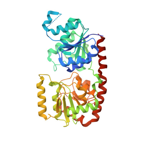
    Formation of the heterocyst envelope polysaccharide (HEP) is a key process for cyanobacterial heterocyst differentiation. The maturation of HEP in Anabaena sp. strain PCC 7120 is controlled by a gene cluster termed HEP island in addition to an operon alr3698-alr3699, which encodes two putative proteins termed Alr3698/HepD and Alr3699/HepE. Here we report the crystal structures of HepE in the apo-form and three complex forms that bind to UDP-glucose (UDPG), UDP&glucose, and UDP, respectively. The overall structure of HepE displays a typical GT-B fold of glycosyltransferases, comprising two separate β/α/β Rossmann-fold domains that form an inter-domain substrate-binding crevice. Structural analyses combined with enzymatic assays indicate that HepE is a glucosyltransferase using UDPG as a sugar donor. Further site-directed mutageneses enable us to assign the key residues that stabilize the sugar donor and putative acceptor. Based on the comparative structural analyses, we propose a putative catalytic cycle of HepE, which undergoes "open-closed-open" conformational changes upon binding to the substrates and release of products. These findings provide structural and catalytic insights into the first enzyme involved in the HEP biosynthesis pathway.


  • Organizational Affiliation
    • Hefei National Laboratory for Physical Sciences at the Microscale and School of Life Sciences, University of Science and Technology of China, Hefei, Anhui 230027, People's Republic of China.

Macromolecule Content 

  • Total Structure Weight: 86.87 kDa 
  • Atom Count: 5,962 
  • Modeled Residue Count: 735 
  • Deposited Residue Count: 776 
  • Unique protein chains: 1

Macromolecules

Find similar proteins by:|  3D Structure
Entity ID: 1
MoleculeChains  Sequence LengthOrganismDetailsImage
Alr3699 proteinA [auth B],
B [auth A]
388Nostoc sp. PCC 7120 = FACHB-418Mutation(s): 0 
Gene Names: alr3699
EC: 2.4.1
UniProt
Find proteins for A0ACD6B985 (Nostoc sp. (strain PCC 7120 / SAG 25.82 / UTEX 2576))
Explore A0ACD6B985 
Go to UniProtKB:  A0ACD6B985
Entity Groups
Sequence Clusters30% Identity50% Identity70% Identity90% Identity95% Identity100% Identity
UniProt GroupA0ACD6B985
Sequence Annotations
Expand
Reference Sequence

Small Molecules

Ligands 4 Unique
IDChains Name / Formula / InChI Key2D Diagram3D Interactions
UDP

Query on UDP


Download:Ideal Coordinates CCD File
D [auth B],
K [auth A]
URIDINE-5'-DIPHOSPHATE
C9 H14 N2 O12 P2
XCCTYIAWTASOJW-XVFCMESISA-N
GLC

Query on GLC


Download:Ideal Coordinates CCD File
C [auth B],
J [auth A]
alpha-D-glucopyranose
C6 H12 O6
WQZGKKKJIJFFOK-DVKNGEFBSA-N
SO4

Query on SO4


Download:Ideal Coordinates CCD File
E [auth B]
F [auth B]
G [auth B]
H [auth B]
I [auth B]
E [auth B],
F [auth B],
G [auth B],
H [auth B],
I [auth B],
L [auth A],
M [auth A],
N [auth A],
O [auth A]
SULFATE ION
O4 S
QAOWNCQODCNURD-UHFFFAOYSA-L
GOL

Query on GOL


Download:Ideal Coordinates CCD File
P [auth A]GLYCEROL
C3 H8 O3
PEDCQBHIVMGVHV-UHFFFAOYSA-N

Experimental Data & Validation

Experimental Data

  • Method: X-RAY DIFFRACTION
  • Resolution: 2.48 Å
  • R-Value Free:  0.267 (Depositor), 0.268 (DCC) 
  • R-Value Work:  0.223 (Depositor), 0.230 (DCC) 
  • R-Value Observed: 0.225 (Depositor) 
Space Group: P 32 2 1
Unit Cell:
Length ( Å )Angle ( ˚ )
a = 130.991α = 90
b = 130.991β = 90
c = 156.327γ = 120
Software Package:
Software NamePurpose
REFMACrefinement
HKL-2000data processing
HKL-2000data scaling
MOLREPmodel building
MOLREPphasing

Structure Validation

View Full Validation Report


Ligand Structure Quality Assessment 


Entry History 

& Funding Information

Deposition Data


Funding OrganizationLocationGrant Number
National Natural Science Foundation of ChinaChina31370757

Revision History  (Full details and data files)

  • Version 1.0: 2016-01-13
    Type: Initial release
  • Version 1.1: 2016-04-13
    Changes: Database references
  • Version 1.2: 2020-07-29
    Type: Remediation
    Reason: Carbohydrate remediation
    Changes: Data collection, Database references, Derived calculations, Structure summary
  • Version 1.3: 2023-11-08
    Changes: Data collection, Database references, Refinement description, Structure summary