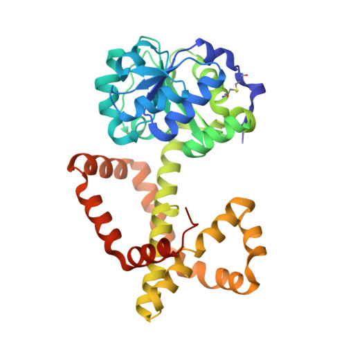
4XIY

Crystal structure of ketol-acid reductoisomerase from Azotobacter


Experimental Data Snapshot

  • Method: X-RAY DIFFRACTION
  • Resolution: 2.50 Å
  • R-Value Free: 0.244 
  • R-Value Work: 0.194 
  • R-Value Observed: 0.196 

Starting Model: experimental
View more details

wwPDB Validation   3D Report Full Report


This is version 1.3 of the entry. See complete history


Literature

Cofactor specificity motifs and the induced fit mechanism in class I ketol-acid reductoisomerases.

Cahn, J.K.Brinkmann-Chen, S.Spatzal, T.Wiig, J.A.Buller, A.R.Einsle, O.Hu, Y.Ribbe, M.W.Arnold, F.H.

(2015) Biochem J 468: 475-484

  • DOI: https://doi.org/10.1042/BJ20150183
  • Primary Citation of Related Structures:  
    4XDY, 4XDZ, 4XEH, 4XIY

  • PubMed Abstract: 

    Although most sequenced members of the industrially important ketol-acid reductoisomerase (KARI) family are class I enzymes, structural studies to date have focused primarily on the class II KARIs, which arose through domain duplication. In the present study, we present five new crystal structures of class I KARIs. These include the first structure of a KARI with a six-residue β2αB (cofactor specificity determining) loop and an NADPH phosphate-binding geometry distinct from that of the seven- and 12-residue loops. We also present the first structures of naturally occurring KARIs that utilize NADH as cofactor. These results show insertions in the specificity loops that confounded previous attempts to classify them according to loop length. Lastly, we explore the conformational changes that occur in class I KARIs upon binding of cofactor and metal ions. The class I KARI structures indicate that the active sites close upon binding NAD(P)H, similar to what is observed in the class II KARIs of rice and spinach and different from the opening of the active site observed in the class II KARI of Escherichia coli. This conformational change involves a decrease in the bending of the helix that runs between the domains and a rearrangement of the nicotinamide-binding site.


  • Organizational Affiliation

    Division of Chemistry and Chemical Engineering, California Institute of Technology, Pasadena, CA 91125, U.S.A.


Macromolecules
Find similar proteins by:  (by identity cutoff)  |  3D Structure
Entity ID: 1
MoleculeChains Sequence LengthOrganismDetailsImage
Ketol-acid reductoisomerase
A, B, C, D
338Azotobacter vinelandii DJMutation(s): 0 
EC: 1.1.1.86
UniProt
Find proteins for C1DFH7 (Azotobacter vinelandii (strain DJ / ATCC BAA-1303))
Explore C1DFH7 
Go to UniProtKB:  C1DFH7
Entity Groups  
Sequence Clusters30% Identity50% Identity70% Identity90% Identity95% Identity100% Identity
UniProt GroupC1DFH7
Sequence Annotations
Expand
  • Reference Sequence
Small Molecules
Ligands 5 Unique
IDChains Name / Formula / InChI Key2D Diagram3D Interactions
PG4
Query on PG4

Download Ideal Coordinates CCD File 
P [auth D]TETRAETHYLENE GLYCOL
C8 H18 O5
UWHCKJMYHZGTIT-UHFFFAOYSA-N
PGE
Query on PGE

Download Ideal Coordinates CCD File 
G [auth A],
J [auth B]
TRIETHYLENE GLYCOL
C6 H14 O4
ZIBGPFATKBEMQZ-UHFFFAOYSA-N
PEG
Query on PEG

Download Ideal Coordinates CCD File 
M [auth C]DI(HYDROXYETHYL)ETHER
C4 H10 O3
MTHSVFCYNBDYFN-UHFFFAOYSA-N
FE
Query on FE

Download Ideal Coordinates CCD File 
F [auth A],
I [auth B],
K [auth C],
O [auth D]
FE (III) ION
Fe
VTLYFUHAOXGGBS-UHFFFAOYSA-N
MG
Query on MG

Download Ideal Coordinates CCD File 
E [auth A],
H [auth B],
L [auth C],
N [auth D]
MAGNESIUM ION
Mg
JLVVSXFLKOJNIY-UHFFFAOYSA-N
Experimental Data & Validation

Experimental Data

  • Method: X-RAY DIFFRACTION
  • Resolution: 2.50 Å
  • R-Value Free: 0.244 
  • R-Value Work: 0.194 
  • R-Value Observed: 0.196 
  • Space Group: P 21 3
Unit Cell:
Length ( Å )Angle ( ˚ )
a = 185.05α = 90
b = 185.05β = 90
c = 185.05γ = 90
Software Package:
Software NamePurpose
REFMACrefinement
MOSFLMdata reduction
SCALAdata scaling
MOLREPphasing
SHELXphasing

Structure Validation

View Full Validation Report


Entry History 

Deposition Data

Revision History  (Full details and data files)

  • Version 1.0: 2015-04-22
    Type: Initial release
  • Version 1.1: 2015-09-02
    Changes: Database references
  • Version 1.2: 2023-09-27
    Changes: Data collection, Database references, Derived calculations, Refinement description
  • Version 1.3: 2024-11-06
    Changes: Structure summary