


Crystal structure of Argininosuccinate synthase from Mycobacterium thermoresistibile in complex with AMPPNP and Arginine
Abendroth, J., Dranow, D.M., Lorimer, D.D., Edwards, T.E.To be published.
Experimental Data Snapshot
Starting Model: experimental
View more details
Entity ID: 1 | |||||
|---|---|---|---|---|---|
| Molecule | Chains | Sequence Length | Organism | Details | Image |
| Argininosuccinate synthase | 408 | Mycolicibacterium thermoresistibile ATCC 19527 | Mutation(s): 0 Gene Names: argG, KEK_01915 EC: 6.3.4.5 | 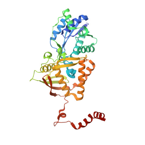 | |
UniProt | |||||
Entity Groups | |||||
| Sequence Clusters | 30% Identity50% Identity70% Identity90% Identity95% Identity100% Identity | ||||
| UniProt Group | G7CBN9 | ||||
Sequence AnnotationsExpand | |||||
Reference Sequence | |||||
| Ligands 4 Unique | |||||
|---|---|---|---|---|---|
| ID | Chains | Name / Formula / InChI Key | 2D Diagram | 3D Interactions | |
| ANP Download:Ideal Coordinates CCD File | C [auth A], K [auth B] | PHOSPHOAMINOPHOSPHONIC ACID-ADENYLATE ESTER C10 H17 N6 O12 P3 PVKSNHVPLWYQGJ-KQYNXXCUSA-N |  | ||
| ARG Download:Ideal Coordinates CCD File | E [auth A], M [auth B] | ARGININE C6 H15 N4 O2 ODKSFYDXXFIFQN-BYPYZUCNSA-O |  | ||
| EDO Download:Ideal Coordinates CCD File | F [auth A] G [auth A] I [auth A] J [auth B] N [auth B] | 1,2-ETHANEDIOL C2 H6 O2 LYCAIKOWRPUZTN-UHFFFAOYSA-N |  | ||
| MG Download:Ideal Coordinates CCD File | D [auth A], H [auth A], L [auth B] | MAGNESIUM ION Mg JLVVSXFLKOJNIY-UHFFFAOYSA-N |  | ||
| Length ( Å ) | Angle ( ˚ ) |
|---|---|
| a = 93.83 | α = 90 |
| b = 143.41 | β = 90 |
| c = 58.03 | γ = 90 |
| Software Name | Purpose |
|---|---|
| XDS | data reduction |
| XSCALE | data scaling |
| PHENIX | refinement |
| Coot | model building |
| PDB_EXTRACT | data extraction |
| PHENIX | phasing |


