4XEC | pdb_00004xec

Staphylococcus aureus Dihydrofolate Reductase complexed with NADPH and 6-ETHYL-5-[(3R)-3-[3-METHOXY-5-(PYRIDIN-4-YL)PHENYL]BUT-1-YN-1-YL]PYRIMIDINE-2,4-DIAMINE (UCP1061)


Experimental Data Snapshot

  • Method: X-RAY DIFFRACTION
  • Resolution: 2.69 Å
  • R-Value Free: 
    0.233 (Depositor), 0.230 (DCC) 
  • R-Value Work: 
    0.156 (Depositor), 0.162 (DCC) 
  • R-Value Observed: 
    0.164 (Depositor) 

Starting Model: experimental
View more details

wwPDB Validation 3D Report Full Report

Validation slider image for 4XEC

Ligand Structure Quality Assessment 


This is version 1.5 of the entry. See complete history

Literature

Nonracemic Antifolates Stereoselectively Recruit Alternate Cofactors and Overcome Resistance in S. aureus.

Keshipeddy, S.Reeve, S.M.Anderson, A.C.Wright, D.L.

(2015) J Am Chem Soc 137: 8983-8990

  • DOI: https://doi.org/10.1021/jacs.5b01442
  • Primary Citation Related Structures: 
    4TU5, 4XEC

  • PubMed Abstract: 

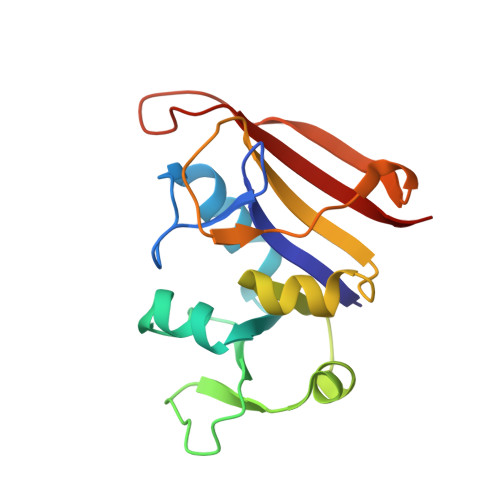
    While antifolates such as Bactrim (trimethoprim-sulfamethoxazole; TMP-SMX) continue to play an important role in treating community-acquired methicillin-resistant Staphylococcus aureus (CA-MRSA), resistance-conferring mutations, specifically F98Y of dihydrofolate reductase (DHFR), have arisen and compromise continued use. In an attempt to extend the lifetime of this important class, we have developed a class of propargyl-linked antifolates (PLAs) that exhibit potent inhibition of the enzyme and bacterial strains. Probing the role of the configuration at the single propargylic stereocenter in these inhibitors required us to develop a new approach to nonracemic 3-aryl-1-butyne building blocks by the pairwise use of asymmetric conjugate addition and aldehyde dehydration protocols. Using this new route, a series of nonracemic PLA inhibitors was prepared and shown to possess potent enzyme inhibition (IC50 values <50 nM), antibacterial effects (several with MIC values <1 μg/mL) and to form stable ternary complexes with both wild-type and resistant mutants. Unexpectedly, crystal structures of a pair of individual enantiomers in the wild-type DHFR revealed that the single change in configuration of the stereocenter drove the selection of an alternative NADPH cofactor, with the minor α-anomer appearing with R-27. Remarkably, this cofactor switching becomes much more prevalent when the F98Y mutation is present. The observation of cofactor site plasticity leads to a postulate for the structural basis of TMP resistance in DHFR and also suggests design strategies that can be used to target these resistant enzymes.


  • Organizational Affiliation
    • Department of Pharmaceutical Sciences, University of Connecticut, 69 North Eagleville Road, Storrs, Connecticut 06269, United States.

Macromolecule Content 

  • Total Structure Weight: 19.19 kDa 
  • Atom Count: 1,398 
  • Modeled Residue Count: 157 
  • Deposited Residue Count: 157 
  • Unique protein chains: 1

Macromolecules

Find similar proteins by:|  3D Structure
Entity ID: 1
MoleculeChains  Sequence LengthOrganismDetailsImage
Dihydrofolate reductaseA [auth X]157Staphylococcus aureusMutation(s): 0 
Gene Names: folA
EC: 1.5.1.3
UniProt
Find proteins for P0A017 (Staphylococcus aureus)
Explore P0A017 
Go to UniProtKB:  P0A017
Entity Groups
Sequence Clusters30% Identity50% Identity70% Identity90% Identity95% Identity100% Identity
UniProt GroupP0A017
Sequence Annotations
Expand
Reference Sequence

Small Molecules

Ligands 3 Unique
IDChains Name / Formula / InChI Key2D Diagram3D Interactions
NDP

Query on NDP


Download:Ideal Coordinates CCD File
D [auth X]NADPH DIHYDRO-NICOTINAMIDE-ADENINE-DINUCLEOTIDE PHOSPHATE
C21 H30 N7 O17 P3
ACFIXJIJDZMPPO-NNYOXOHSSA-N
06U

Query on 06U


Download:Ideal Coordinates CCD File
B [auth X]6-ethyl-5-{(3R)-3-[3-methoxy-5-(pyridin-4-yl)phenyl]but-1-yn-1-yl}pyrimidine-2,4-diamine
C22 H23 N5 O
KEPLBUUTAQCZOE-AWEZNQCLSA-N
ACT

Query on ACT


Download:Ideal Coordinates CCD File
C [auth X]ACETATE ION
C2 H3 O2
QTBSBXVTEAMEQO-UHFFFAOYSA-M

Experimental Data & Validation

Experimental Data

  • Method: X-RAY DIFFRACTION
  • Resolution: 2.69 Å
  • R-Value Free:  0.233 (Depositor), 0.230 (DCC) 
  • R-Value Work:  0.156 (Depositor), 0.162 (DCC) 
  • R-Value Observed: 0.164 (Depositor) 
Space Group: P 61 2 2
Unit Cell:
Length ( Å )Angle ( ˚ )
a = 78.902α = 90
b = 78.902β = 90
c = 108.118γ = 120
Software Package:
Software NamePurpose
PHENIXrefinement
HKL-2000data reduction
HKL-2000data scaling
PHENIXphasing

Structure Validation

View Full Validation Report


Ligand Structure Quality Assessment 


Entry History 

& Funding Information

Deposition Data


Funding OrganizationLocationGrant Number
National Institutes of Health/National Institute Of Allergy and Infectious Diseases (NIH/NIAID)United StatesAI111957

Revision History  (Full details and data files)

  • Version 1.0: 2015-07-22
    Type: Initial release
  • Version 1.1: 2015-08-05
    Changes: Database references
  • Version 1.2: 2017-09-20
    Changes: Author supporting evidence, Derived calculations
  • Version 1.3: 2018-01-31
    Changes: Experimental preparation
  • Version 1.4: 2019-12-11
    Changes: Author supporting evidence
  • Version 1.5: 2023-09-27
    Changes: Data collection, Database references, Refinement description