4XB4 | pdb_00004xb4

Structure of the N-terminal domain of OCP binding canthaxanthin


Experimental Data Snapshot

  • Method: X-RAY DIFFRACTION
  • Resolution: 1.54 Å
  • R-Value Free: 
    0.160 (Depositor), 0.160 (DCC) 
  • R-Value Work: 
    0.147 (Depositor), 0.151 (DCC) 
  • R-Value Observed: 
    0.148 (Depositor) 

Starting Model: experimental
View more details

wwPDB Validation 3D Report Full Report

Validation slider image for 4XB4

Ligand Structure Quality Assessment 


This is version 1.4 of the entry. See complete history

Literature

PHOTOSYNTHESIS. A 12 angstrom carotenoid translocation in a photoswitch associated with cyanobacterial photoprotection.

Leverenz, R.L.Sutter, M.Wilson, A.Gupta, S.Thurotte, A.Bourcier de Carbon, C.Petzold, C.J.Ralston, C.Perreau, F.Kirilovsky, D.Kerfeld, C.A.

(2015) Science 348: 1463-1466

  • DOI: https://doi.org/10.1126/science.aaa7234
  • Primary Citation Related Structures: 
    4XB4, 4XB5

  • PubMed Abstract: 

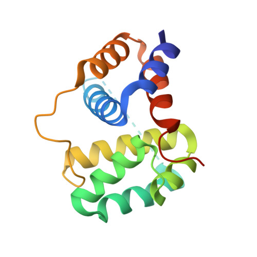
    Pigment-protein and pigment-pigment interactions are of fundamental importance to the light-harvesting and photoprotective functions essential to oxygenic photosynthesis. The orange carotenoid protein (OCP) functions as both a sensor of light and effector of photoprotective energy dissipation in cyanobacteria. We report the atomic-resolution structure of an active form of the OCP consisting of the N-terminal domain and a single noncovalently bound carotenoid pigment. The crystal structure, combined with additional solution-state structural data, reveals that OCP photoactivation is accompanied by a 12 angstrom translocation of the pigment within the protein and a reconfiguration of carotenoid-protein interactions. Our results identify the origin of the photochromic changes in the OCP triggered by light and reveal the structural determinants required for interaction with the light-harvesting antenna during photoprotection.


  • Organizational Affiliation
    • MSU-DOE Plant Research Laboratory, Michigan State University, East Lansing, MI 48824, USA.

Macromolecule Content 

  • Total Structure Weight: 34.34 kDa 
  • Atom Count: 2,644 
  • Modeled Residue Count: 282 
  • Deposited Residue Count: 304 
  • Unique protein chains: 1

Macromolecules

Find similar proteins by:|  3D Structure
Entity ID: 1
MoleculeChains  Sequence LengthOrganismDetailsImage
Orange carotenoid-binding protein
A, B
152Synechocystis sp. PCC 6803 substr. KazusaMutation(s): 0 
Gene Names: slr1963
UniProt
Find proteins for P74102 (Synechocystis sp. (strain ATCC 27184 / PCC 6803 / Kazusa))
Explore P74102 
Go to UniProtKB:  P74102
Entity Groups
Sequence Clusters30% Identity50% Identity70% Identity90% Identity95% Identity100% Identity
UniProt GroupP74102
Sequence Annotations
Expand
Reference Sequence

Experimental Data & Validation

Experimental Data

  • Method: X-RAY DIFFRACTION
  • Resolution: 1.54 Å
  • R-Value Free:  0.160 (Depositor), 0.160 (DCC) 
  • R-Value Work:  0.147 (Depositor), 0.151 (DCC) 
  • R-Value Observed: 0.148 (Depositor) 
Space Group: P 1 21 1
Unit Cell:
Length ( Å )Angle ( ˚ )
a = 51.251α = 90
b = 64.112β = 114.89
c = 53.634γ = 90
Software Package:
Software NamePurpose
PHENIXrefinement
SCALAdata scaling
XDSdata scaling
PHASERphasing

Structure Validation

View Full Validation Report


Ligand Structure Quality Assessment 


Entry History 

Deposition Data

Revision History  (Full details and data files)

  • Version 1.0: 2015-07-15
    Type: Initial release
  • Version 1.1: 2015-09-09
    Changes: Data collection
  • Version 1.2: 2015-12-30
    Changes: Non-polymer description
  • Version 1.3: 2017-11-22
    Changes: Derived calculations, Refinement description
  • Version 1.4: 2023-09-27
    Changes: Data collection, Database references, Refinement description