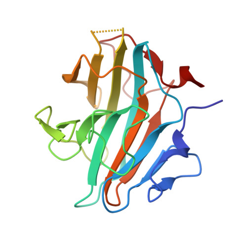
4X8P | pdb_00004x8p

Crystal structure of Ash2L SPRY domain in complex with RbBP5


Experimental Data Snapshot

  • Method: X-RAY DIFFRACTION
  • Resolution: 2.20 Å
  • R-Value Free: 
    0.271 (Depositor), 0.261 (DCC) 
  • R-Value Work: 
    0.220 (Depositor), 0.207 (DCC) 
  • R-Value Observed: 
    0.222 (Depositor) 

wwPDB Validation 3D Report Full Report

Validation slider image for 4X8P

This is version 1.1 of the entry. See complete history. 

Literature

A phosphorylation switch on RbBP5 regulates histone H3 Lys4 methylation.

Zhang, P., Chaturvedi, C.P., Tremblay, V., Cramet, M., Brunzelle, J.S., Skiniotis, G., Brand, M., Shilatifard, A., Couture, J.F.

(2015) Genes Dev 29: 123-128

  • DOI: https://doi.org/10.1101/gad.254870.114
  • Primary Citation Related Structures: 
    4X8N, 4X8P

  • PubMed Abstract: 

    The methyltransferase activity of the trithorax group (TrxG) protein MLL1 found within its COMPASS (complex associated with SET1)-like complex is allosterically regulated by a four-subunit complex composed of WDR5, RbBP5, Ash2L, and DPY30 (also referred to as WRAD). We report structural evidence showing that in WRAD, a concave surface of the Ash2L SPIa and ryanodine receptor (SPRY) domain binds to a cluster of acidic residues, referred to as the D/E box, in RbBP5. Mutational analysis shows that residues forming the Ash2L/RbBP5 interface are important for heterodimer formation, stimulation of MLL1 catalytic activity, and erythroid cell terminal differentiation. We also demonstrate that a phosphorylation switch on RbBP5 stimulates WRAD complex formation and significantly increases KMT2 (lysine [K] methyltransferase 2) enzyme methylation rates. Overall, our findings provide structural insights into the assembly of the WRAD complex and point to a novel regulatory mechanism controlling the activity of the KMT2/COMPASS family of lysine methyltransferases.


  • Organizational Affiliation: 
    • Department of Biochemistry, Microbiology, and Immunology, Ottawa Institute of Systems Biology, University of Ottawa, Ottawa, Ontario K1H 8M5, Canada;

Macromolecule Content 

  • Total Structure Weight: 22.08 kDa 
  • Atom Count: 1,610 
  • Modeled Residue Count: 188 
  • Deposited Residue Count: 194 
  • Unique protein chains: 2

Macromolecules

Find similar proteins by:|  3D Structure
Entity ID: 1
MoleculeChains  Sequence LengthOrganismDetailsImage
Set1/Ash2 histone methyltransferase complex subunit ASH2,Set1/Ash2 histone methyltransferase complex subunit ASH2182Homo sapiensMutation(s): 0 
Gene Names: ASH2L, ASH2L1
UniProt & NIH Common Fund Data Resources
Find proteins for Q9UBL3 (Homo sapiens)
Explore Q9UBL3 
Go to UniProtKB:  Q9UBL3
PHAROS:  Q9UBL3
GTEx:  ENSG00000129691 
Entity Groups
Sequence Clusters30% Identity50% Identity70% Identity90% Identity95% Identity100% Identity
UniProt GroupQ9UBL3
Sequence Annotations
Expand
Reference Sequence
Find similar proteins by:  Sequence   |   3D Structure  
Entity ID: 2
MoleculeChains  Sequence LengthOrganismDetailsImage
Retinoblastoma-binding protein 512Homo sapiensMutation(s): 0 
UniProt & NIH Common Fund Data Resources
Find proteins for Q15291 (Homo sapiens)
Explore Q15291 
Go to UniProtKB:  Q15291
PHAROS:  Q15291
GTEx:  ENSG00000117222 
Entity Groups
Sequence Clusters30% Identity50% Identity70% Identity90% Identity95% Identity100% Identity
UniProt GroupQ15291
Sequence Annotations
Expand
Reference Sequence

Small Molecules

Ligands 1 Unique
IDChains Name / Formula / InChI Key2D Diagram3D Interactions
GOL

Query on GOL


Download:Ideal Coordinates CCD File
C [auth B]GLYCEROL
C3 H8 O3
PEDCQBHIVMGVHV-UHFFFAOYSA-N

Experimental Data & Validation

Experimental Data

  • Method: X-RAY DIFFRACTION
  • Resolution: 2.20 Å
  • R-Value Free:  0.271 (Depositor), 0.261 (DCC) 
  • R-Value Work:  0.220 (Depositor), 0.207 (DCC) 
  • R-Value Observed: 0.222 (Depositor) 
Space Group: P 21 21 21
Unit Cell:
Length ( Å )Angle ( ˚ )
a = 51.5α = 90
b = 59.1β = 90
c = 70.8γ = 90
Software Package:
Software NamePurpose
BUSTERrefinement
d*TREKdata reduction
d*TREKdata scaling
PHASERphasing

Structure Validation

View Full Validation Report


Entry History 

Deposition Data

Revision History  (Full details and data files)

  • Version 1.0: 2015-01-28
    Type: Initial release
  • Version 1.1: 2024-02-28
    Changes: Data collection, Database references, Derived calculations, Source and taxonomy