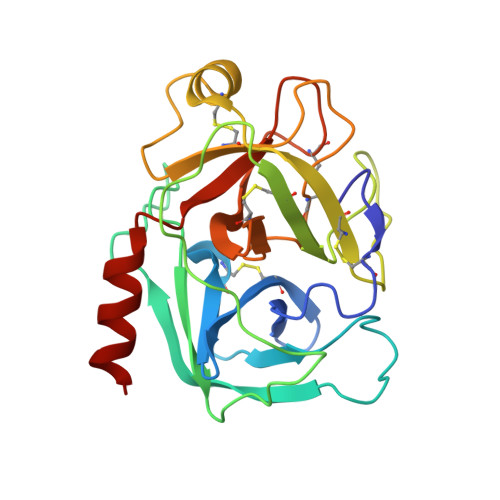
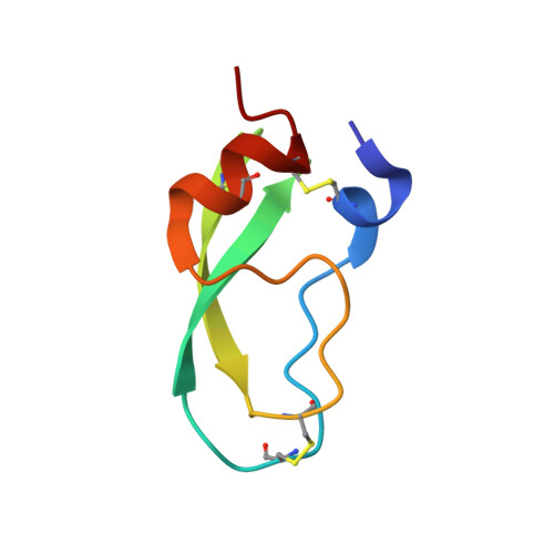
4WWY

human cationic trypsin G193R mutant in complex with bovine pancreatic trypsin inhibitor


Experimental Data Snapshot

  • Method: X-RAY DIFFRACTION
  • Resolution: 1.70 Å
  • R-Value Free: 0.207 
  • R-Value Work: 0.171 
  • R-Value Observed: 0.173 

Starting Model: experimental
View more details

wwPDB Validation   3D Report Full Report


This is version 1.7 of the entry. See complete history


Literature

Mesotrypsin Has Evolved Four Unique Residues to Cleave Trypsin Inhibitors as Substrates.

Alloy, A.P.Kayode, O.Wang, R.Hockla, A.Soares, A.S.Radisky, E.S.

(2015) J Biol Chem 290: 21523-21535

  • DOI: https://doi.org/10.1074/jbc.M115.662429
  • Primary Citation of Related Structures:  
    4WWY, 4WXV

  • PubMed Abstract: 

    Human mesotrypsin is highly homologous to other mammalian trypsins, and yet it is functionally unique in possessing resistance to inhibition by canonical serine protease inhibitors and in cleaving these inhibitors as preferred substrates. Arg-193 and Ser-39 have been identified as contributors to the inhibitor resistance and cleavage capability of mesotrypsin, but it is not known whether these residues fully account for the unusual properties of mesotrypsin. Here, we use human cationic trypsin as a template for engineering a gain of catalytic function, assessing mutants containing mesotrypsin-like mutations for resistance to inhibition by bovine pancreatic trypsin inhibitor (BPTI) and amyloid precursor protein Kunitz protease inhibitor (APPI), and for the ability to hydrolyze these inhibitors as substrates. We find that Arg-193 and Ser-39 are sufficient to confer mesotrypsin-like resistance to inhibition; however, compared with mesotrypsin, the trypsin-Y39S/G193R double mutant remains 10-fold slower at hydrolyzing BPTI and 2.5-fold slower at hydrolyzing APPI. We identify two additional residues in mesotrypsin, Lys-74 and Asp-97, which in concert with Arg-193 and Ser-39 confer the full catalytic capability of mesotrypsin for proteolysis of BPTI and APPI. Novel crystal structures of trypsin mutants in complex with BPTI suggest that these four residues function cooperatively to favor conformational dynamics that assist in dissociation of cleaved inhibitors. Our results reveal that efficient inhibitor cleavage is a complex capability to which at least four spatially separated residues of mesotrypsin contribute. These findings suggest that inhibitor cleavage represents a functional adaptation of mesotrypsin that may have evolved in response to positive selection pressure.


  • Organizational Affiliation

    From the Department of Cancer Biology, Mayo Clinic Comprehensive Cancer Center, Jacksonville, Florida 32224 and.


Macromolecules
Find similar proteins by:  (by identity cutoff)  |  3D Structure
Entity ID: 1
MoleculeChains Sequence LengthOrganismDetailsImage
Trypsin-1
A, B
224Homo sapiensMutation(s): 2 
Gene Names: PRSS1TRP1TRY1TRYP1
EC: 3.4.21.4
UniProt & NIH Common Fund Data Resources
Find proteins for P07477 (Homo sapiens)
Explore P07477 
Go to UniProtKB:  P07477
PHAROS:  P07477
GTEx:  ENSG00000204983 
Entity Groups  
Sequence Clusters30% Identity50% Identity70% Identity90% Identity95% Identity100% Identity
UniProt GroupP07477
Sequence Annotations
Expand
  • Reference Sequence
Find similar proteins by:  (by identity cutoff)  |  3D Structure
Entity ID: 2
MoleculeChains Sequence LengthOrganismDetailsImage
Pancreatic trypsin inhibitorC,
D [auth I]
58Bos taurusMutation(s): 0 
UniProt
Find proteins for P00974 (Bos taurus)
Explore P00974 
Go to UniProtKB:  P00974
Entity Groups  
Sequence Clusters30% Identity50% Identity70% Identity90% Identity95% Identity100% Identity
UniProt GroupP00974
Sequence Annotations
Expand
  • Reference Sequence
Small Molecules
Ligands 2 Unique
IDChains Name / Formula / InChI Key2D Diagram3D Interactions
SO4
Query on SO4

Download Ideal Coordinates CCD File 
E [auth A]
F [auth A]
G [auth A]
I [auth B]
J [auth C]
E [auth A],
F [auth A],
G [auth A],
I [auth B],
J [auth C],
K [auth C],
L [auth I],
M [auth I]
SULFATE ION
O4 S
QAOWNCQODCNURD-UHFFFAOYSA-L
CA
Query on CA

Download Ideal Coordinates CCD File 
H [auth B]CALCIUM ION
Ca
BHPQYMZQTOCNFJ-UHFFFAOYSA-N
Experimental Data & Validation

Experimental Data

  • Method: X-RAY DIFFRACTION
  • Resolution: 1.70 Å
  • R-Value Free: 0.207 
  • R-Value Work: 0.171 
  • R-Value Observed: 0.173 
  • Space Group: P 1 21 1
Unit Cell:
Length ( Å )Angle ( ˚ )
a = 52.952α = 90
b = 63.227β = 94.74
c = 90.535γ = 90
Software Package:
Software NamePurpose
REFMACrefinement
HKL-2000data reduction
PDB_EXTRACTdata extraction
HKL-2000data scaling

Structure Validation

View Full Validation Report


Entry History & Funding Information

Deposition Data


Funding OrganizationLocationGrant Number
National Institutes of Health/National Cancer Institute (NIH/NCI)United StatesR01CA154387

Revision History  (Full details and data files)

  • Version 1.0: 2015-07-22
    Type: Initial release
  • Version 1.1: 2015-07-29
    Changes: Database references
  • Version 1.2: 2015-08-19
    Changes: Experimental preparation
  • Version 1.3: 2015-09-09
    Changes: Database references
  • Version 1.4: 2017-09-13
    Changes: Author supporting evidence, Database references, Derived calculations
  • Version 1.5: 2019-12-04
    Changes: Author supporting evidence
  • Version 1.6: 2023-09-27
    Changes: Data collection, Database references, Refinement description
  • Version 1.7: 2024-11-20
    Changes: Structure summary